Cutaneous Schwannoma in a Cow
DOI:
https://doi.org/10.24070/bjvp.1983-0246.005015Keywords:
Schwannoma, histopathology, immunohistochemistry (IHC),, skin, cattleAbstract
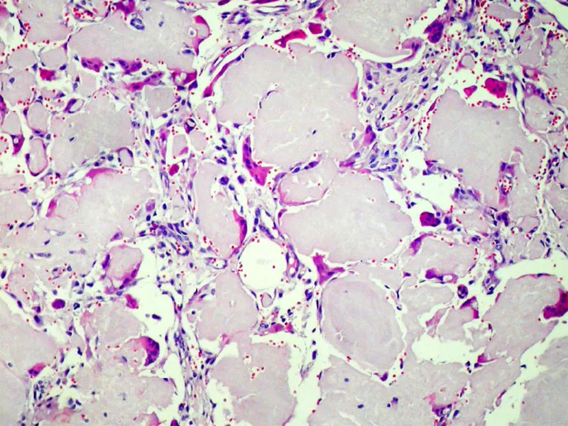
Polioencephalomalacia (PEM) of ruminants is a complex disease. The term indicates a morphological diagnosis where severe cortical neuronal necrosis results in softening of cerebralSchwannoma is a peripheral nerve sheath tumor (PNST), commonly found as a spindle cell tumor of autonomic nerves and rarely involving the skin of cattle. The present report describes the histopathology and immunohistochemistry features of a localized (solitary) benign PNST with final diagnosis of cutaneous schwannoma in a 4-year-old female Holstein bovine. The dome-shaped, well circumscribed, firm, non-smooth surfaced mass was composed of spindle-shaped cells arranged predominantly in interlacing fascicles or streams with a moderate to strong intervening collagenous stroma. Histopathologic changes included typical hypocellular areas with pale scant eosinophilic cytoplasm (Antoni B pattern) similar to myxomatous tissue, and hypercellular areas with deeply eosinophilic cytoplasm (non-typical Antoni A pattern) without nuclear palisading or Verocay bodies formation. Immunohistochemical reactions for S-100 protein, a Schwann cell marker, and vimentin were strong in the neoplastic cells. Other markers as desmin, neuron specific enolase (NSE), CD34, and p53 were all negative. It was concluded that concurrent evaluation of both histological and immunohistochemical features are required for the final diagnosis of schwannomas in domestic animals. grey matter. Initially though as a single disease caused by thiamine deficiency, it is currently believed that PEM is caused by different etiological agents through different pathogenic mechanisms or trough a single pathogenic mechanism triggered by different agents. In this paper the putative cases and pathogenesis of PEM in ruminants are critically reviewed and discussed. Also reviewed are the epidemiology, clinical signs, gross and histological findings and methods of diagnosis of cases of PEM described in ruminants in Brazil