Comparative Analysis of Histopathological Alterations and Immunohistochemistry in Cattle for Diagnosis of Rabies
DOI:
https://doi.org/10.24070/bjvp.1983-0246.006001Keywords:
cattle, histopathology, immunohistochemistry, rabiesAbstract
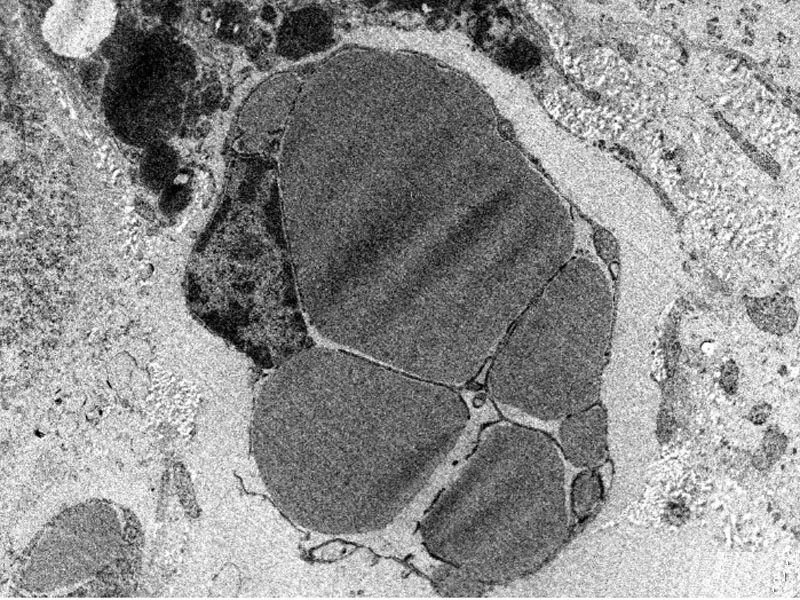
Comparative analysis of histopathological alterations and immunohistochemistry was performed on 15 brain samples of cow. In the present study, brain tissues were positive in 9 cases (60.0%), using polyclonal antiserum by immunohistochemistry and 7 cases (46.60%) were found positive for rabies by demonstration of Negri bodies. Hundred neurons per case were observed for Negri bodies; number of Negri bodies in positive neurons was counted and a comparison of IHC and histopathology was done. Number of neurons positive for rabies virus antigen per 100 neurons (900 neurons) by IHC were more (665) than H& E staining (344) and average number of Negri bodies per neuron detected by IHC were more (2.97) than histopathology (1.52), therefore, IHC was found to be more sensitive than histopathology.


