
Sebaceous epithelioma in a ferret (Mustela putorius furo)
DOI:
https://doi.org/10.24070/bjvp.1983-0246.001014Keywords:
Sebaceous Epithelioma, Ferret, neoplasia, pathology, diseases of Ferrets, Mustela putorius furoAbstract
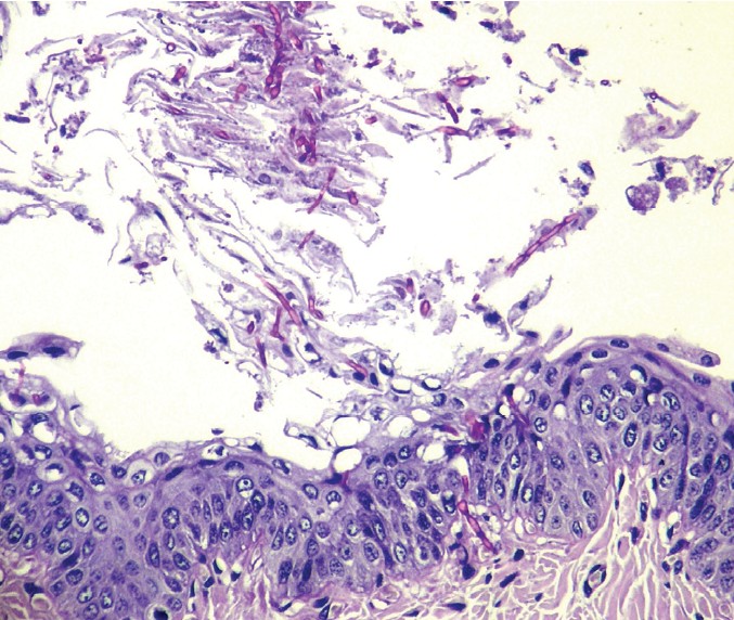
An eight years-old male ferret presented a ventral abdominal nodular mass near the inguinal region. On clinical examination, a warty nodular mass in the abdomen was detected and the ferret was submitted to excisional biopsy by surgical procedure. At histopathology, adenomatous structures with preponderance of basophilic reserve cells, some sebocytes and showing squamous differentiation were observed. The diagnosis of sebaceous epithelioma was established based on clinical presentation and histopathologic findings. The animal recovered and at the moment no complication was reported.
Downloads
Published
2008-11-30
Issue
Section
Articles
How to Cite
Bonel-Raposo, J., Silveira, M. F., Gamba, C. de O., Spader, M. B., Guim, T. N., Schuch, I. D., Ladeira, R. S., & Fernandes, C. G. (2008). Sebaceous epithelioma in a ferret (Mustela putorius furo). Brazilian Journal of Veterinary Pathology, 1(2), 70-72. https://doi.org/10.24070/bjvp.1983-0246.001014