Morphological and Immunophenotipical Characterization of Murine Mammary Carcinoma 4t1
DOI:
https://doi.org/10.24070/bjvp.1983-0246.007018Keywords:
mouse, mammary gland, 4T1 cells and immunohistochemistryAbstract
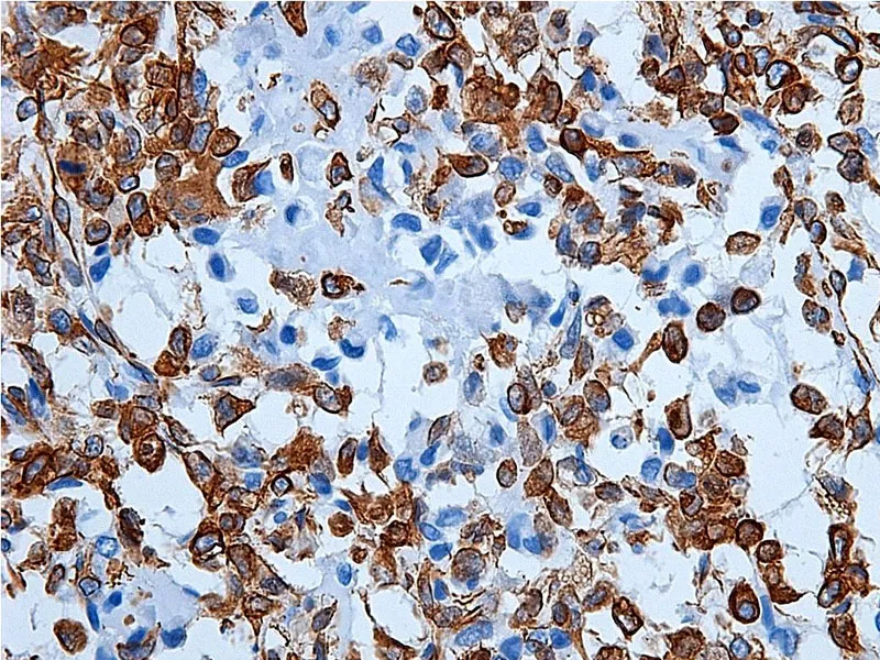
The 4T1 murine mammary carcinoma is an experimental model widely used in assessing and better understanding of tumor biology. It is a highly tumorigenic cell line and invasive, where metastases are observed in various organs. This study aims to describe morphological and immunophenotipical aspects of 4T1 mammary carcinoma in mice Balb/c with the aid of the immunohistochemistry. Tissues were fixed in formalin and processed using the routine paraffin inclusion technique. Histologic sections (4 μm) were stained through Hematoxylin-Eosin techniques for morphologic assessments. For immunohistochemical study, we used a panel of 9 (nine) antibodies: hormone receptors, receptors for cell proliferation, cytokeratins, vimentin, growth factor receptor and markers of blood vessels. Morphologically, the 4T1 murine mammary carcinoma shows malignant epithelial proliferation in solid arrangement, with pleomorphic cells and high mitotic index. In immunohistochemical analysis was determined a positivity for hormone receptors, cytokeratin AE1/AE3, receptors of cell proliferation and markers of blood vessels. There was a negative for vimentin, cytokeratin 5/6, cytokeratin 34βE12 and growth factor receptor.The results show that the characteristics observed in this model are similar to some types of breast cancers found in women like poorly differentiated invasive ductal carcinoma. Thus, the immunophenotypic characterization of mammary carcinoma 4T1 allows a better understanding of the model to the study of new anticancer therapies.