Renal encephalopathy due to acute renal failure in a goat

Authors

  • Fábio B. Rosa Programa de Pós-Graduação em Medicina Veterinária, Centro de Ciências Rurais (CCR), Universidade Federal de Santa Maria (UFSM) Author
  • Mara I. B. Rubin Programa de Pós-Graduação em Medicina Veterinária, Centro de Ciências Rurais (CCR), Universidade Federal de Santa Maria (UFSM) Author
  • Paula V. Leal Laboratório de Anatomia Patológica - Faculdade de Medicina Veterinária e Zootecnia (FAMEZ), Universidade Federal de Mato Grosso do Sul (UFMS) Author
  • Tessie B. Martins Laboratório de Anatomia Patológica - Faculdade de Medicina Veterinária e Zootecnia (FAMEZ), Universidade Federal de Mato Grosso do Sul (UFMS) Author
  • Danilo C. Gomes Laboratório de Anatomia Patológica - Faculdade de Medicina Veterinária e Zootecnia (FAMEZ), Universidade Federal de Mato Grosso do Sul (UFMS) Author
  • Marcelo A. Araújo Setor de Clínica Médica e Cirúrgica de Grandes Animais (FAMEZ/UFSM). Author
  • Ricardo A. A. Lemos Laboratório de Anatomia Patológica - Faculdade de Medicina Veterinária e Zootecnia (FAMEZ), Universidade Federal de Mato Grosso do Sul (UFMS) Author
  • Claudio S. L. Barros Laboratório de Anatomia Patológica - Faculdade de Medicina Veterinária e Zootecnia (FAMEZ), Universidade Federal de Mato Grosso do Sul (UFMS), Author

Keywords:

diseases of goats, renal encephalopathy, status spongiosus, acute renal failure, sulfadiazine

Abstract

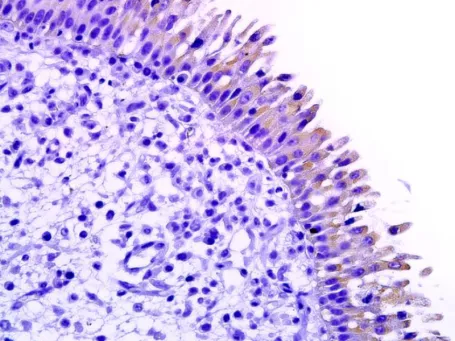
Renal encephalopathy was diagnosed in a 7-month-old male goat with a history of diarrhea and dehydration due to Eimeria sp. infection. The goat was treated with sulfadiazine before developing central nervous system (CNS) signs characterized by severe anorexia, salivation, tremors, inability to stand and depression. Biochemical parameters revealed high levels of blood urea nitrogen (BUN) and creatinine, 263.6 mg/dL and 2.9 mg/dL respectively. No gross pathological changes were observed at necropsy. Histopathological examination of the brain revealed large irregular empty spaces (status spongiosus) in the white matter of the brainstem, cerebellum, thalamus, basal nuclei and in the interface of white and grey matter in the cerebrum. There was severe multifocal renal tubular necrosis characterized by abundant deposits of basophilic granular material, frequently forming crystals that replaced the lost tubular epithelial cells and filled the lumina. The clinical-pathologic findings support to a diagnosis of encephalopathy due to acute renal failure.

Downloads

Published

2015-11-30

Issue

Section

Artigos

How to Cite

Rosa, F. B., Rubin, M. I. B., Leal, P. V., Martins, T. B., Gomes, D. C., Araújo, M. A., Lemos, R. A. A., & Barros, C. S. L. (2015). Renal encephalopathy due to acute renal failure in a goat. Brazilian Journal of Veterinary Pathology, 8(3), 112-115. https://bjvp.org.br/bjvp/article/view/206