Frequency, pathology and electron microscopy of dromedary camel viral fibro-papilloma in Sudan
DOI:
https://doi.org/10.24070/bjvp.1983-0246.009007Keywords:
camels, epidemiology, pathology, electron microscopy, viral fibro-papillomaAbstract
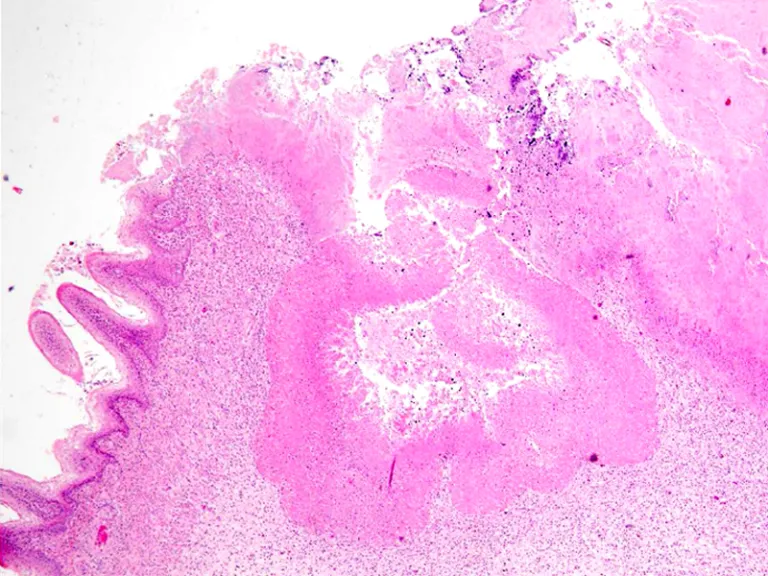
Sixteen different herds comprising 1803 camels were surveyed between August 2009 and January 2012 for presence of Camel Papillomatosis. Outbreaks of the disease were observed in two different areas. The first outbreak occurred in Al-Qutaynah locality, about 83 Km south of Khartoum and the second in Al-Fashagah locality about 410 Km south east of Khartoum. Fifty three camels were found to be affected with papillomatosis, with a total morbidity rate of 2.9%. All affected animals were 3-24 months old in addition to 2 females aged four and five years old. Cases of Camel Papillomatosis were recorded in January, July, August and October. The skin lesions were dark grey or white keratinized fissured raised masses, some of which were pedunculated. They showed various shapes: round, oval, cauliflower, horn shape, flat or dome shape and measured on average about 8.8 X 7.5 X 7.1 mm. Warts occurred mostly in head and face but other sites (limbs, ventral abdomen, sternum and tail) were also involved. Twenty Five cases were analyzed histopathologically, in which sections were typical for fibropapilloma characterized by multiple papillary proliferations covered with keratinized epithelium, with down growth of rete ridges. Acanthosis with karyopyknosis and cytoplasmic vacuolations in stratum spinosum cells and hyperkeratosis were seen together with subepithelial fibrosis. No inclusions could be detected in squamous cells. Out of 19 samples investigated immunohistochemically for papillomavirus antigens, 10 samples were found positive. Using transmission electron microscopy, aggregates of papillomavirus virions were found in the nuclei of the stratum granulosum in one sample.