Primary diffuse mammary hemangiosarcoma in a female dog

Authors

  • Antônio F. Leis Filho Department of Animal Reproduction and Veterinary Radiology, São Paulo State University – UNESP, Botucatu, São Paulo, Brazil Author
  • Patricia F. Lainetti Department of Animal Reproduction and Veterinary Radiology, São Paulo State University – UNESP, Botucatu, São Paulo, Brazil Author
  • Marina A. Cyrino Department of Animal Reproduction and Veterinary Radiology, São Paulo State University – UNESP, Botucatu, São Paulo, Brazil Author
  • Priscila E. Kobayashi Department of Veterinary Clinic, São Paulo State University – UNESP, Botucatu, São Paulo, Brazil Author
  • Camila D. Costa Department of Veterinary Clinic, São Paulo State University – UNESP, Botucatu, São Paulo, Brazil Author
  • Laíza S. Camargo Department of Animal Reproduction and Veterinary Radiology, São Paulo State University – UNESP, Botucatu, São Paulo, Brazil Author
  • Fabiana F. Souza Department of Animal Reproduction and Veterinary Radiology, São Paulo State University – UNESP, Botucatu, São Paulo, Brazil Author
  • Renée Laufer-Amorim Department of Veterinary Clinic, São Paulo State University – UNESP, Botucatu, São Paulo, Brazil Author
  • Carlos E. Fonseca-Alves Department of Veterinary Clinic, São Paulo State University – UNESP, Botucatu, São Paulo, Brazil Author

DOI:

https://doi.org/10.24070/bjvp.1983-0246.v11i3p102-107

Keywords:

dog, mammary neoplasia, angiosarcoma, endothelial cells, neoplasia

Abstract

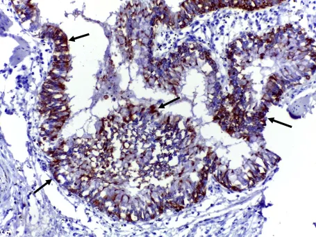
In veterinary medicine, primary mammary hemangiosarcoma is a very rare disease and there was no previous report describing the disease in dogs. Herein, we describe necropsy findings of a female dog, which presented a diffuse mammary hemangiosarcoma affecting the mammary gland. A diffuse irregular plaque was found in all mammary gland, involving the whole mammary gland tissue. The histopathological evaluation revealed neoplastic cells with an ovoid to spindle shaped basophilic and numerous atypical neoplastic tumor cells forming vascular structures. There was no glandular proliferation and no changes in the superficial dermis and no neoplastic emboli in the lymph vessels. Immunohistochemistry for vimentin, pan-cytokeratin, CK8/18 and CD31 was conducted. Positive expression was found to the pan-cytokeratin and CK8/18 by remaining epithelial cells confirmed the luminal mammary origin. Vimentin and CD31 positive expression by neoplastic cells confirmed the endothelial origin of the neoplasia. The histopathological and immunohistochemical findings supported the diagnosis of a primary mammary hemangiosarcoma.

Downloads

Published

2018-11-30

Issue

Section

Artigos

How to Cite

Leis Filho, A. F., Lainetti, P. F., Cyrino, M. A., Kobayashi, P. E., Costa, C. D., Camargo, L. S., Souza, F. F., Laufer-Amorim, R., & Fonseca-Alves, C. E. (2018). Primary diffuse mammary hemangiosarcoma in a female dog. Brazilian Journal of Veterinary Pathology, 11(3), 102-107. https://doi.org/10.24070/bjvp.1983-0246.v11i3p102-107