Canine distemper virus, atypical Toxoplasma gondii, and Neospora caninum co-infection, in a dog with neurological signs from Argentina
DOI:
https://doi.org/10.24070/bjvp.1983-0246.v12i3p101-105Keywords:
Toxoplasma gondii, Neospora caninum, canine distemper virus, genotypingAbstract
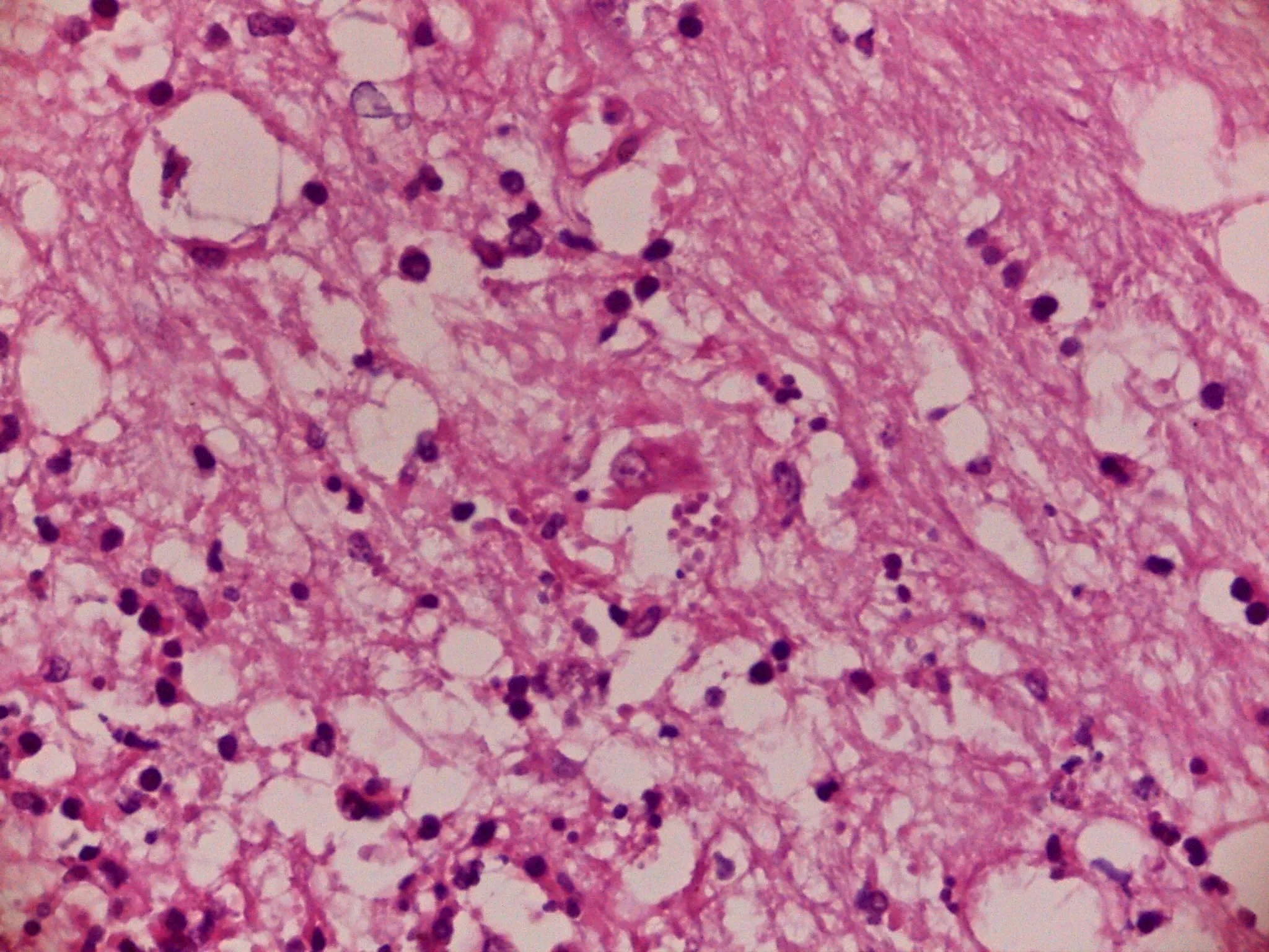
The occurrence of combined infections by Toxoplasma gondii, Neospora caninum and Canine distemper virus (CDV) in domestic dogs and wildlife animals has not been frequently reported, and the histopathological findings were not exhaustively described. The objective of this study was to report a co-infection of CDV, T. gondii and N. caninum in a dog with neurological signs, as well as the molecular characterization of the protozoa involved. A young street dog was rescued with neurological clinical signs and died spontaneously. A complete necropsy was performed. Tissues were collected and fixed for histopathological evaluation. Additionally, sections of the central nervous system (CNS) and heart were assayed by immunohistochemistry (IHQ) for T. gondii and N. caninum. Sample of brain tissue was analyzed by PCR and nPCR-RFLP for T. gondii genotyping. Spleen was used for detection of CDV by RT-PCR. Gross lesions were not observed, with the exception of the lung. Microscopically, a severe necrosuppurative meningoencephalitis with vasculitis, tachyzoites and bradyzoites of T. gondii and N. caninum were found. Demyelination was also evident, associated with eosinophilic intranuclear inclusion bodies within astrocytes. CDV was PCR positive while both parasites were presented PCR and IHQ positive results. Molecular characterization of T. gondii was reported as atypical #14 (likely). To our knowledge, this is the first report of genetical identification of T. gondii obtained from the brain of a naturally infected dog in Argentina. The results emphasize the importance of different techniques as diagnostic tools to enhance the detection of causative agents in cases of fatal encephalitis.