Use of Giemsa staining for the immunohistochemical counterstaining in canine melanomas: an “old and forgotten” method
DOI:
https://doi.org/10.24070/bjvp.1983-0246.v13i1p17-20Keywords:
Giemsa, immunodetection, melanin, pigmented neoplasmAbstract
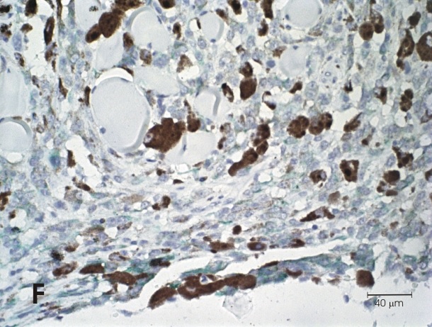
Melanoma is a neoplasm originating from melanocytes and represents 7% of skin tumors and the most common in oral cavity of dogs. Melanoma may present melanocytic pigment in its cytoplasm in varying quantity and its characterization by immunohistochemistry (IHC) is challenging due to the use of chromogen diaminobenzidine (DAB) which itself produces a brown product what makes difficult to distinguish from melanin pigment in this technique. To demonstrate a reliable technique, the use of the Giemsa counterstaining was performed in the IHC of melanomas with different degrees of pigmentation for different cytoplasmic, membrane and nuclear markers. The modification in the IHC technique by the counterstaining of Giemsa allows observable differences under the microscope between melanic pigment (in a blue-green stain), while the DAB chromogen will be observed in brown. With this technique, the prognostic and predictive interpretation of markers in canine melanomas may be more reliable in definitions of clinical behaviors and in experimental analyzes.