Bilateral canine seminoma with ocular metastasis
histochemical and immunohistochemical characterization
DOI:
https://doi.org/10.24070/bjvp.1983-0246.v13i1p57-61Keywords:
C-KIT, Ki-67, inhibin-α, immunohistochemistry, neoplasia, dogAbstract
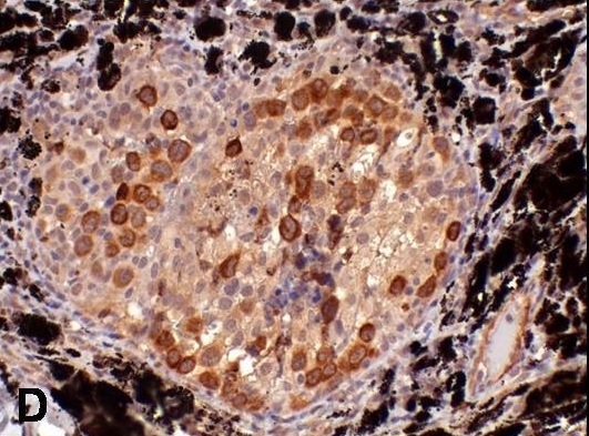
A 10-year old male dog was examined due to a buphthalmia in the left eye and a nodule in the two testicles. Due to the limited resources of the owner and loss of visual acuity of the patient, the enucleation and castration were chosen as treatment. Microscopic analysis of the testicular tissue revealed neoplastic germ cells. Morphologically, neoplastic cells were characterized by distinct cell borders, scarce and eosinophilic cytoplasm, large round nucleus, with thick chromatin and a prominent nucleolus. Binucleated and multinucleated neoplastic cells were also frequently observed. In 10 high power fields (400x), 62 typical and atypical mitosis were counted. Similar neoplastic cells were identified within the vessels of the retina, sclera and in the sub-epithelial conjunctive tissue of the eyelid. The neoplastic cells observed in the testicle and in the eye were positive for PAS. By immunochemistry technique was identified an intense immunostaining of the neoplastic cells for Vimentin and Ki-67 in both testicular and ocular tissue. While, discrete immunoreactivity was identified to c-KIT from the neoplastic cells in both organs. Based on morphological, histochemical and immunohistochemical analysis, it was possible to characterize the ocular lesion as seminoma metastasis.