Intravascular lymphoma in a dog

case report

Authors

  • Rafael R. M. E. Costa Discente do curso de graduação em Medicina Veterinária, Faculdade de Ciências Agrárias e Veterinárias (FCAV-Unesp Jaboticabal), Jaboticabal, SP, Brasil. Author
  • Paulo E. B. Martinelli Residente do Programa de Residência Profissional na área da Saúde – Medicina Veterinária e Saúde, FCAV- UNESP, Jaboticabal, SP, Brasil. Author
  • Isabela F. S. Perossi Residente do Programa de Residência Profissional na área da Saúde – Medicina Veterinária e Saúde, FCAV- UNESP, Jaboticabal, SP, Brasil. Author
  • Leticia Bonato Residente do Programa de Residência Profissional na área da Saúde – Medicina Veterinária e Saúde, FCAV- UNESP, Jaboticabal, SP, Brasil. Author
  • João Victor B. Silva Discente do Centro Universitário Moura Lacerda, Ribeirão Preto, SP, Brasil Author
  • Bruna R. I. Nascimento Discente do Centro Universitário Moura Lacerda, Ribeirão Preto, SP, Brasil Author
  • Paulo H. L. Bertolo Discente do Programa de Pós-graduação em Medicina Veterinária, FCAV- UNESP, Jaboticabal, SP, Brasil. Author
  • Gabriela P. Lima Discente do Programa de Pós-graduação em Medicina Veterinária, FCAV- UNESP, Jaboticabal, SP, Brasil. Author
  • Rosemeri O. Vasconcelos Docente do Departamento de Patologia Veterinária, FCAV – Unesp Jaboticabal, Jaboticabal, SP, Brasil Author

DOI:

https://doi.org/10.24070/bjvp.1983-0246.v13i2p545-548

Keywords:

encephalopathy, lymphoid neoplasm, immunohistochemistry

Abstract

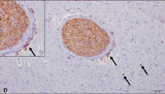
Intravascular lymphoma is characterized by being a malignant neoplasm of extranodal T or B lymphocytes with exclusive proliferation within the vascular lumen, particularly of small vessels. The clinical signs vary due to the involvement of several organs, mainly the central nervous system. The diagnosis is difficult because many of the tests performed are not conclusive and, therefore, necropsy is the most efficient way to identify this tumor. This case report aims to describe the anatomopathological findings of a case of intravascular lymphoma in a mixed breed, 8 years old dog who presented neurological signs and was submitted to a necroscopic examination with clinical suspicion of granulomatous meningoencephalitis. The necropsy findings were not specific, but the presence of intravascular neoplastic lymphocytes in the brain, spleen, adrenal gland and stomach was verified by microscopy. These cells were positive for the CD3 antibody by immunohistochemistry, confirming the T lymphocyte phenotype. This neoplasm should be considered in the diagnosis of encephalopathies in dogs.

Downloads

Published

2020-07-30

Issue

Section

Artigos

How to Cite

Costa, R. R. M. E., Martinelli, P. E. B., Perossi, I. F. S., Bonato, L., Silva, J. V. B., Nascimento, B. R. I., Bertolo, P. H. L., Lima, G. P., & Vasconcelos, R. O. (2020). Intravascular lymphoma in a dog: case report. Brazilian Journal of Veterinary Pathology, 13(2), 545-548. https://doi.org/10.24070/bjvp.1983-0246.v13i2p545-548