Structural description of sea turtle fibropapilloma
DOI:
https://doi.org/10.24070/bjvp.1983-0246.v13i3p585-591Keywords:
fibropapillomatosis, Chelonia mydas, marine diseases, ectoparasitesAbstract
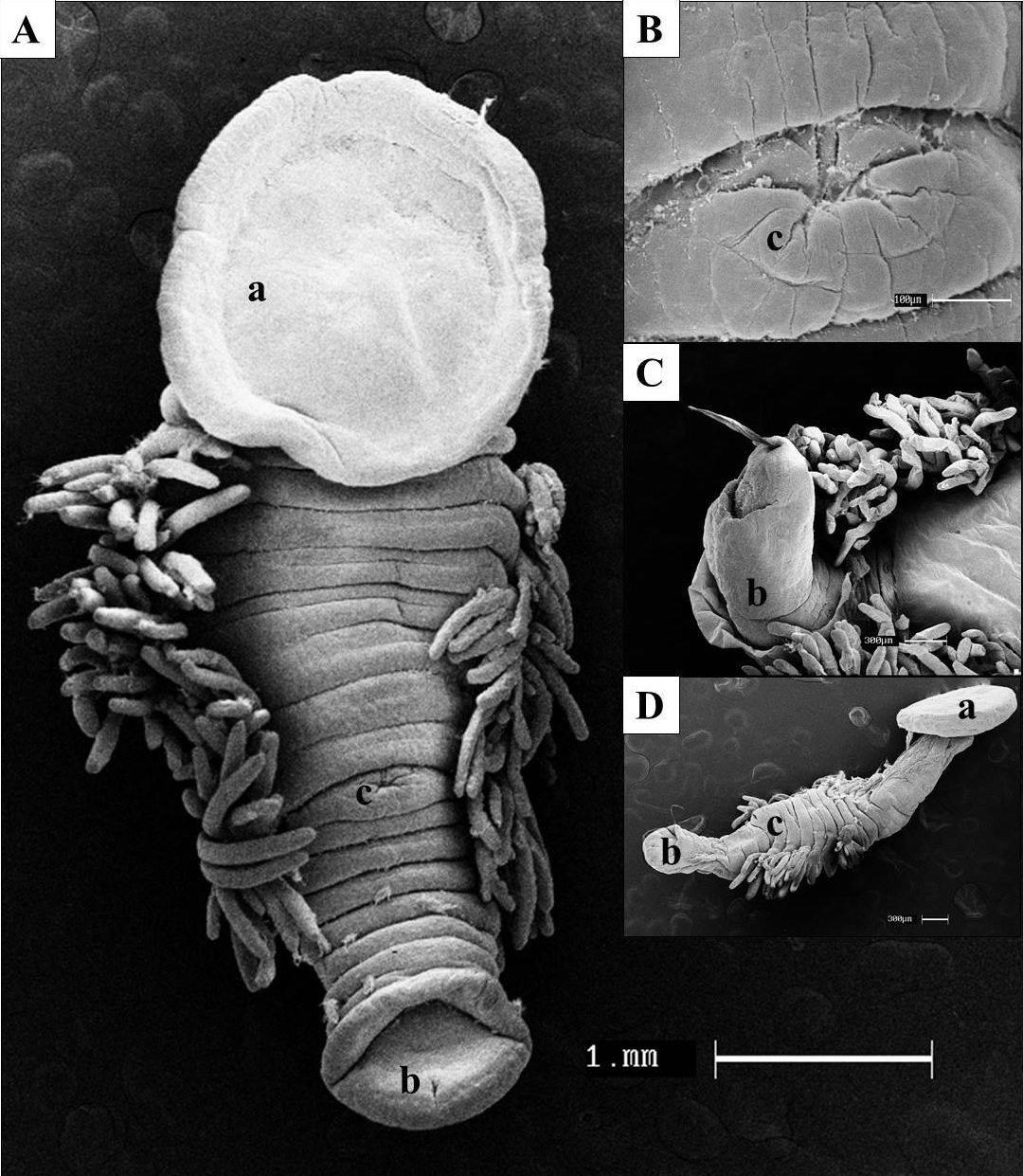
Fibropapillomatosis is a neoplastic disease that affects sea turtles. It is characterized by multiple papillomas, fibropapillomas and cutaneous and/or visceral fibromas. Although its etiology has not been fully elucidated, it is known that there is a strong involvement of an alpha - herpesvirus, but the influence of other factors such as parasites, genetics, chemical carcinogens, contaminants, immunosuppression and ultraviolet radiation may be important in the disease, being pointed out as one of the main causes of a reduction in the green turtle population. Thus, the objective of this article was to describe the morphology of cutaneous fibropapillomas found in specimens of the green turtle (Chelonia mydas), using light and scanning electron microscopy in order to contribute to the mechanism of tumor formation. Microscopically, it presented hyperplastic stromal proliferation and epidermal proliferation with hyperkeratosis. The bulky mass was coated with keratin, with some keratinocyte invaginations, that allowed the keratin to infiltrate from the epidermis into the dermis, forming large keratinized circular spirals. Another fact that we observed was the influence of the inflammation of the tumors caused by ectoparasites.


