Cecal dilatation and distension in a Holstein calf
DOI:
https://doi.org/10.24070/bjvp.1983-0246.v14i1p29-32Keywords:
peritonitis, thrombi, ultrasonographyAbstract
Cecal dilatation and distention is an important disorder in early lactation dairy cows, however, reports describing the
anatomical pathology findings of this condition are scarce in the literature. Etiopathogenesis of cecal dilatation and distention is
often attributed to high concentrate feeds, but there is also evidence of myoelectrical dysfunction contributing to its occurrence.
Diagnosis is often made based on physical exam findings, with the contribution of ancillary exams. This paper aims to describe a
case of cecal dilatation with clinical, laboratorial and pathology findings of a 5-month-old Holstein calf that presented abdominal
distension, positive succussion of the right flank and mild dehydration. Clinical pathology findings included neutrophilic
leukocytosis with regenerative left shift and elevated ruminal chloride. Ultrasonographic examination of the right abdomen
showed distended and hypomotile intestinal loops. Despite that, due to the patient’s age, which prevented rectal palpation, and
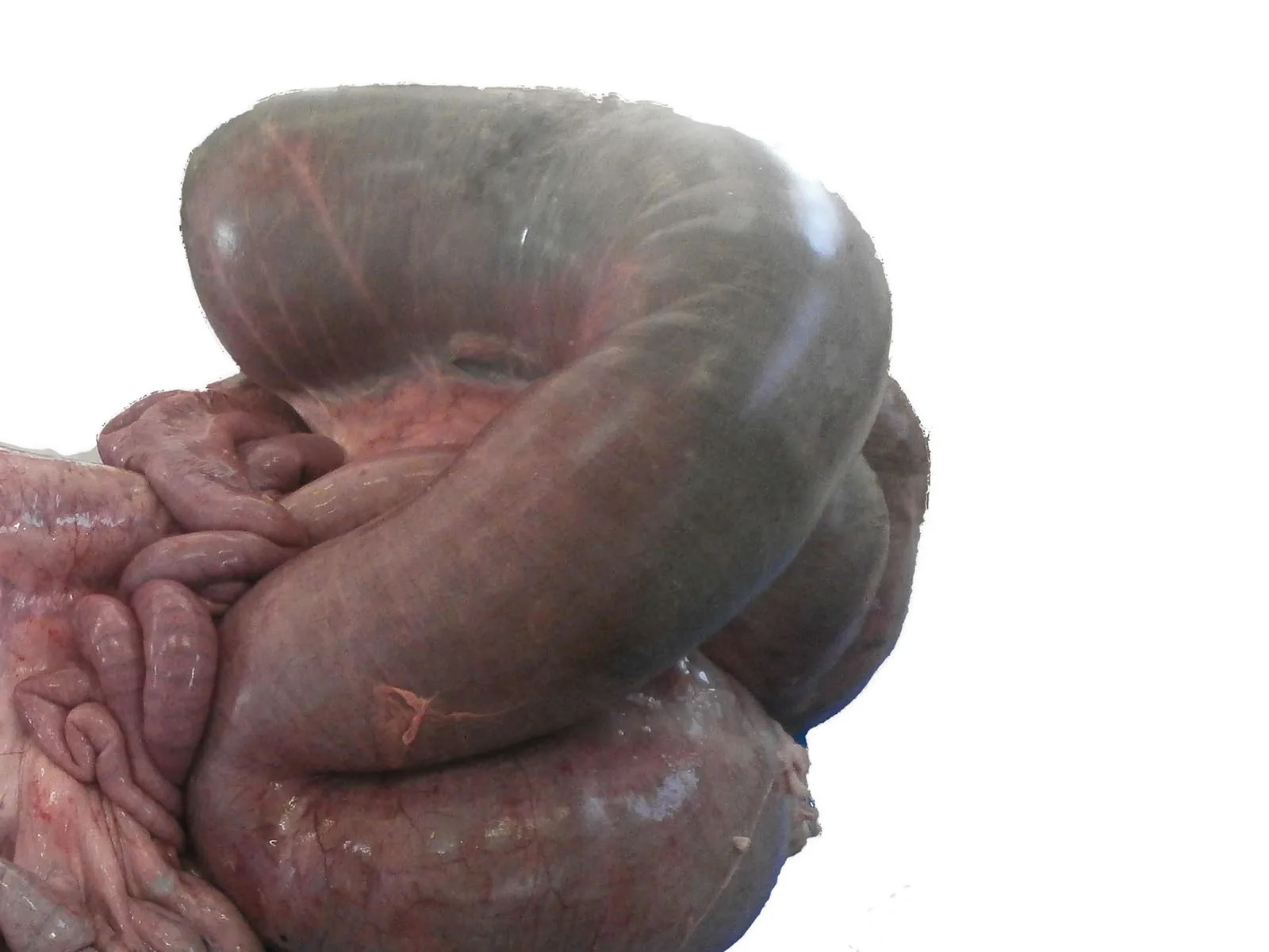
lack of some characteristic clinical and clinical pathology findings, diagnosis was only possible post mortem. Macroscopical and
microscopical findings demonstrated cecum dilatation with edema, hemorrhage and thrombi. Despite being well known by large
animal clinics, anatomical pathologists must be aware of this condition.