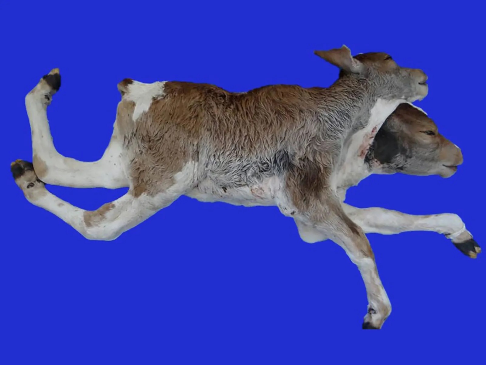
A dicephalus calf
anatomical, pathological and radiographic aspects
DOI:
https://doi.org/10.24070/bjvp.1983-0246.v14i1p33-39Keywords:
congenital malformation, Siamese twins, radiography, ruminant.Abstract
Congenital malformations are morphofunctional abnormalities of tissues and organs that can occur during embryonic
or fetal development in all animal species. Among these, dicephalus is characterized by the development of an individual with
two heads and two necks, due to the total duplication of facial, cranial, and brain structures. Reports of dicephalus in cattle are
scarce and do not normally emphasize radiographic and bone anatomy characteristics. The objective was to describe a case of
a stillborn dicephalus calf. The duplication of the head, brain, neck, and two thoracic vertebral columns, isolated from each
other, with 13 vertebrae each was verified radiographically. There were 13 pairs of ribs, the ones on the right side articulated
with the thoracic spine on the right and the left ones with the spine on the left. Caudally at T13, there was only one lumbar
spine, sacral and coccygeal. In the ventrodorsal projection, L1, L2, L3, and L6 had the shape of a butterfly (suggestive of
hemivertebrae). At necropsy, in addition to craniocervical and spinal morphological changes, collapsed lungs, duplication of
the heart with anastomosis between the aortic arches of the hearts, and duplication of the upper digestive tract were observed.
Additionally, there was arthrogryposis of the pelvic limbs. Corpse maceration, followed by the skeletal assembly, showed the
bone changes previously observed and confirmed the suspicion of hemivertebrae.