Pathology of severe urolithiasis in a flock of egg-laying hens
DOI:
https://doi.org/10.24070/bjvp.1983-0246.v14i1p40-45Keywords:
chickens, histopathology, layers, kidneys, urolithsAbstract
Fourteen, 31-week-old Lohmann white layers from a flock of 30,000 chickens had a history of apathy, and a drop in egg
production. Clinical signs were observed in approximately 40% of the flock, and lasted for three months. Fourteen hens were
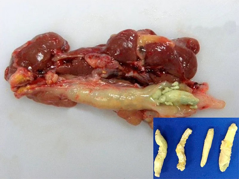
euthanized for post-mortem examinations. Macroscopic findings included marked atrophy and loss of renal lobes along with
compensatory renal hypertrophy of the contralateral lobe. Ureters were markedly dilated and filled with mucus and/or with molded
white to yellow-grey uroliths that obliterated the lumen. At histopathology, the uroliths inside ureters and tubules were composed
of concentrically arranged mineralized concretions, as well as urates associated with heterophilic infiltrations and epithelial
hyperplasia. Renal parenchyma adjacent to obstructed ureters was compressed with tubules replaced by fibrous tissue. Multifocal
interstitial lymphocytic nephritis, proteinuria and membranoproliferative glomerulonephritis were also found. Heterophilic and
caseous ureteritis associated with numerous Gram-positive coccoid bacteria occurred in three chickens. Immunohistochemistry
for avian coronavirus was negative. This negative result along with the case history indicated that water restriction was the most
likely cause of mortality. This condition resulted in significant economic loss for this farmer.