Metastatic calcification and granulomatous grastroenteritis associated to Pythium insidiosum in a dog
DOI:
https://doi.org/10.24070/bjvp.1983-0246.v14i1p50-55Keywords:
oomycete, gastric phytiosis, granuloma, necroscopic analysis, dogAbstract
Pythiosis is a granulomatous process of which the oomycete Pythium insidiosum is its etiological agent. It can affect
animals and humans alike and its infection occurs when free zoospores in the water get in contact with the target tissues and
encyst. The disease often occurs in tropical places with abundance of water and aquatic plants that host the fungus. Dogs
infection is predominantly gastric with granuloma formations in the stomach and intestine with progressive signs of vomiting,
weight loss and diarrhea. In this case report, we described clinical, surgical, necroscopic and histopathological findings of a
one year and two months old, male boxer that presented clinical signs of anorexia and persistent vomiting. It was noticed on
ultrasound examination an increase in stomach and intestine thickness. Laparotomy confirmed a mass affecting the gastric
wall which, an incision biopsy, showed an abundant fibrous tissue associated with granulomatous reaction that was surrounded
by tubuliform structures. Due to clinical complications, euthanasia was performed and in necroscopic examination a markedly
increased stomach and duodenum was observed. An; histological examination of this areas it was observed that they contained
granulation tissue with giant cells and epithelioids macrophages around necrosed areas associated with lymphocytes infiltrate.
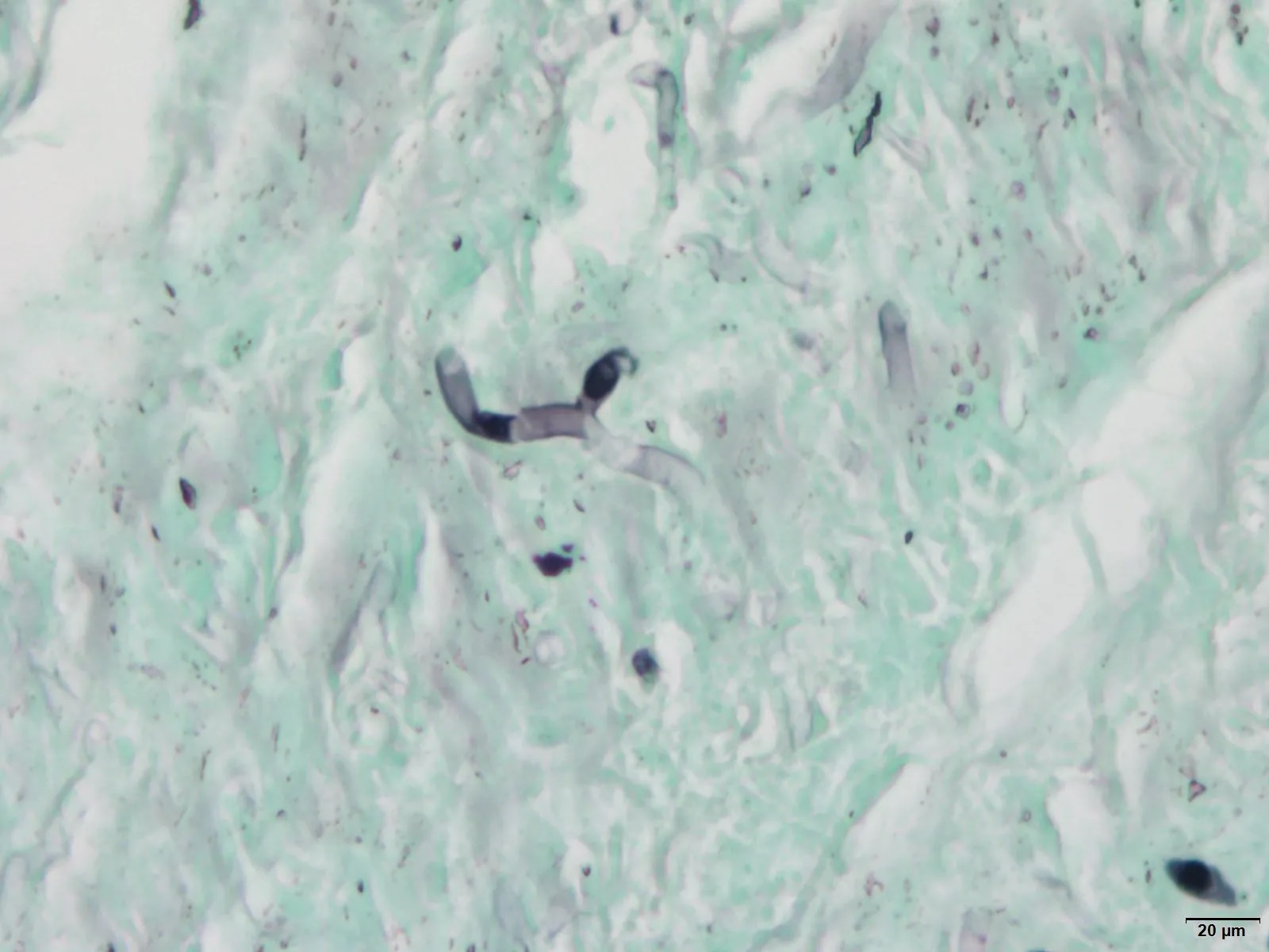
Also, it was possible to observe tubuliform structures by the Grocott-Gomori’s Methenamine Silver (GMS) stain, this finding
is compatible with the agent Pythium insidiosum. Therefore, this presumptive identification was confirmed by PCR analysis
which amplicon had 97.83% similarity with current available genomic sequence of P. insidiosum.