Diaphragmatic hernia and unilateral renal agenesis in a crab-eating fox (Cerdocyon thous)
DOI:
https://doi.org/10.24070/bjvp.1983-0246.v14i1p61-65Keywords:
Wildlife, wild canid, respiratory distress, ultrasonography, radiographyAbstract
An approximately 3-month-old crab-eating fox (Cerdocyon thous) was found by environmental authorities in the State
of Paraiba, Northeastern Brazil and referred to a wildlife care center. The fox was presenting respiratory distress and it was
referred to the Veterinary Hospital of the Federal University of Paraiba (UFPB) for ancillary testing. Abdominal and thoracic
ultrasound and radiographies were performed. These imaging tests indicated the fox had a possible diaphragmatic hernia and
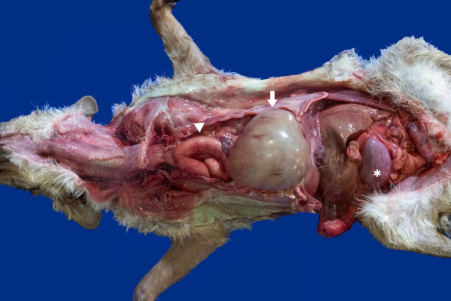
ectopic kidney. The imageology results were confirmed on necropsy, which revealed a postero-lateral focal discontinuity of the
dorsal aspect of the diaphragmatic muscle with protrusion of the gastrointestinal tract into the thoracic cavity. The stomach and
intestinal loops were filled with gas and obliterated the visualization of the heart and lungs. Additionally, only the right kidney
was found, and no vestigial left kidney was identified. Congenital diaphragmatic hernias are not commonly observed in wildlife
but should be considered as a potential differential diagnosis for acute onset of respiratory distress in young carnivores.