Correlation of Clinical, Histopathological and Histomorphometric Features of Canine Soft Tissue Sarcomas
DOI:
https://doi.org/10.24070/bjvp.1983-0246.v14i3p151-158Keywords:
oncology, dog, nucleus-cytoplasm ratio, microvessel densityAbstract
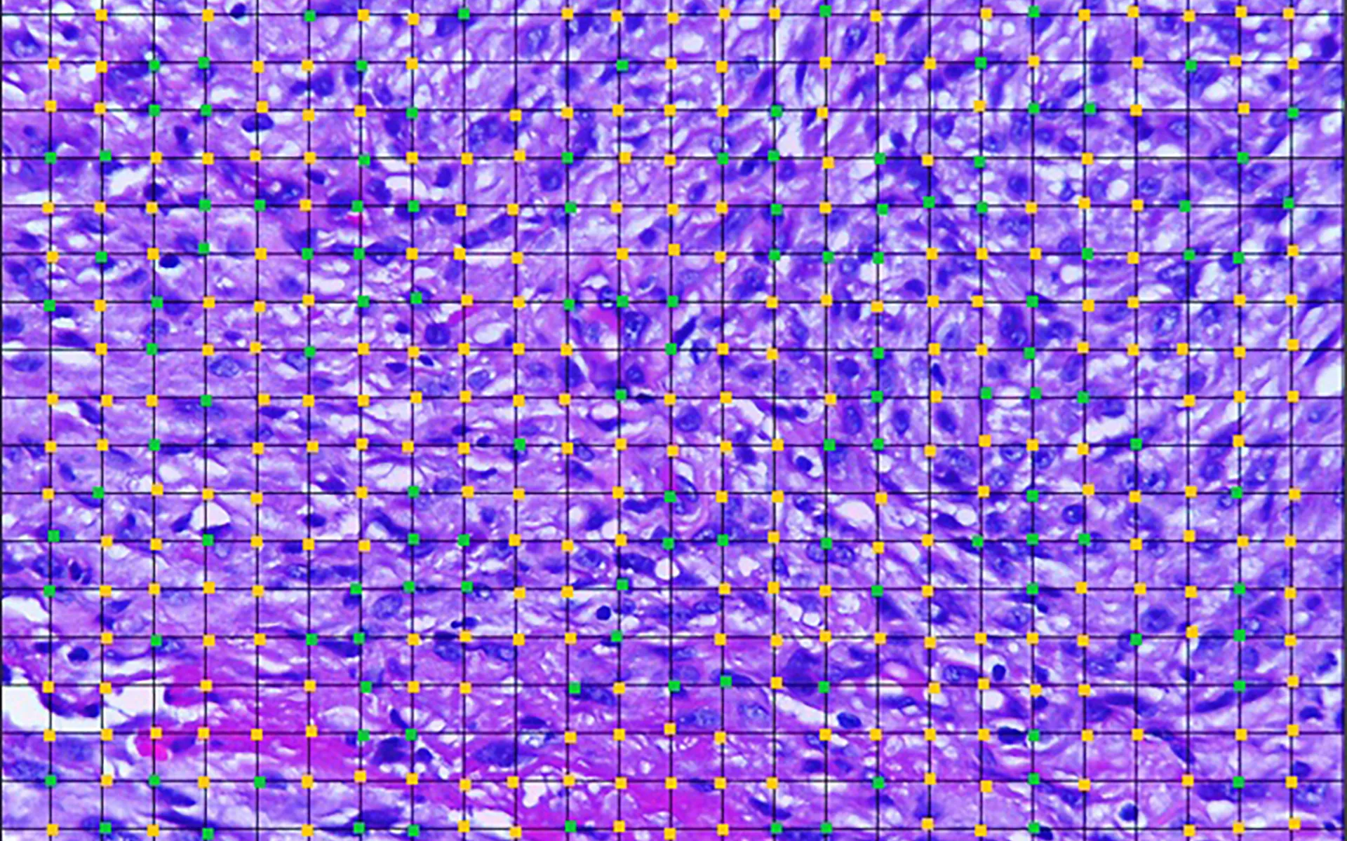
Soft-tissue sarcomas (STS) represent a heterogeneous group of tumours with similar histological characteristics and biological behaviour. This study aimed to describe the correlation between clinical, histopathological and histomorphometric features of STS in dogs. Medical records were reviewed to identify all dogs in which an STS was diagnosed between 2006- 2017. Thirty cases were included, and tumour samples and medical records were recovered. Most of the dogs were mixed breed (40%) and 80% of the STS were located in the subcutaneous connective tissue. Histopathological classification showed that undifferentiated sarcoma (17%) and peripheral nerve sheath tumour (30%) were the most common STS. Grade I STS were obtained in 50% of cases (15/30), and grade II or III tumours compromised 43% (13/30) and 7% (2/30) respectively. The mitotic index ranged from zero to 26 (5.8 ± 7.5). Increased nucleus:cytoplasm ratio was moderately associated with higher tumour grade (p = 0.05; rS = 0.361) and mitotic index (p = 0.05; rS = 0.355), while the number of microvessels was positively correlated with degree of differentiation (p = 0.05; rS = 0.362) and nuclear pleomorphism (p = 0.036; rS = 0.384). Histomorphometry proved to be useful in the evaluation of STS, representing an additional tool correlated with wellestablished prognostic factors (histopathological grade, degree of differentiation, nuclear pleomorphism).