Choroid plexus papilloma in a free ranging eared dove (Zenaida auriculata)

Authors

  • Pedro Enrique Navas-Suárez Divisão de Patologia, Instituto Adolfo Lutz (IAL) Author
  • Ticiana Brasil Ervedosa Divisão de Patologia, Instituto Adolfo Lutz (IAL) Author
  • Celso di Loreto Divisão de Patologia, Instituto Adolfo Lutz (IAL) Author
  • Rodrigo Albergaria Ressio Divisão de Patologia, Instituto Adolfo Lutz (IAL) Author
  • Isis Paixão de Jesus Divisão de Patologia, Instituto Adolfo Lutz (IAL) Author
  • Mariana Morgado Hereny da Silva Centro de Manejo e Conservação de Animais Silvestres - CEMACAS, São Paulo, Brasil. Author
  • Mayra Hespanhol Frediani Centro de Manejo e Conservação de Animais Silvestres - CEMACAS, São Paulo, Brasil. Author
  • Juliana Mariotti Guerra Laboratorio de Patologia Comparada de Animais Selvagens – LAPCOM, Departamento de Patologia, Faculdade de Medicina Veterinária e Zootecnia, Universidade de São Paulo Author
  • Natália C.C. de A. Fernandes Divisão de Patologia, Instituto Adolfo Lutz (IAL), Av. Dr. Arnaldo, 355, São Paulo, SP, Brasil. Author

DOI:

https://doi.org/10.24070/bjvp.1983-0246.v14i3p188-190

Keywords:

Neoplasms, Biodiversity, Wildlife, Choroid plexus tumors

Abstract

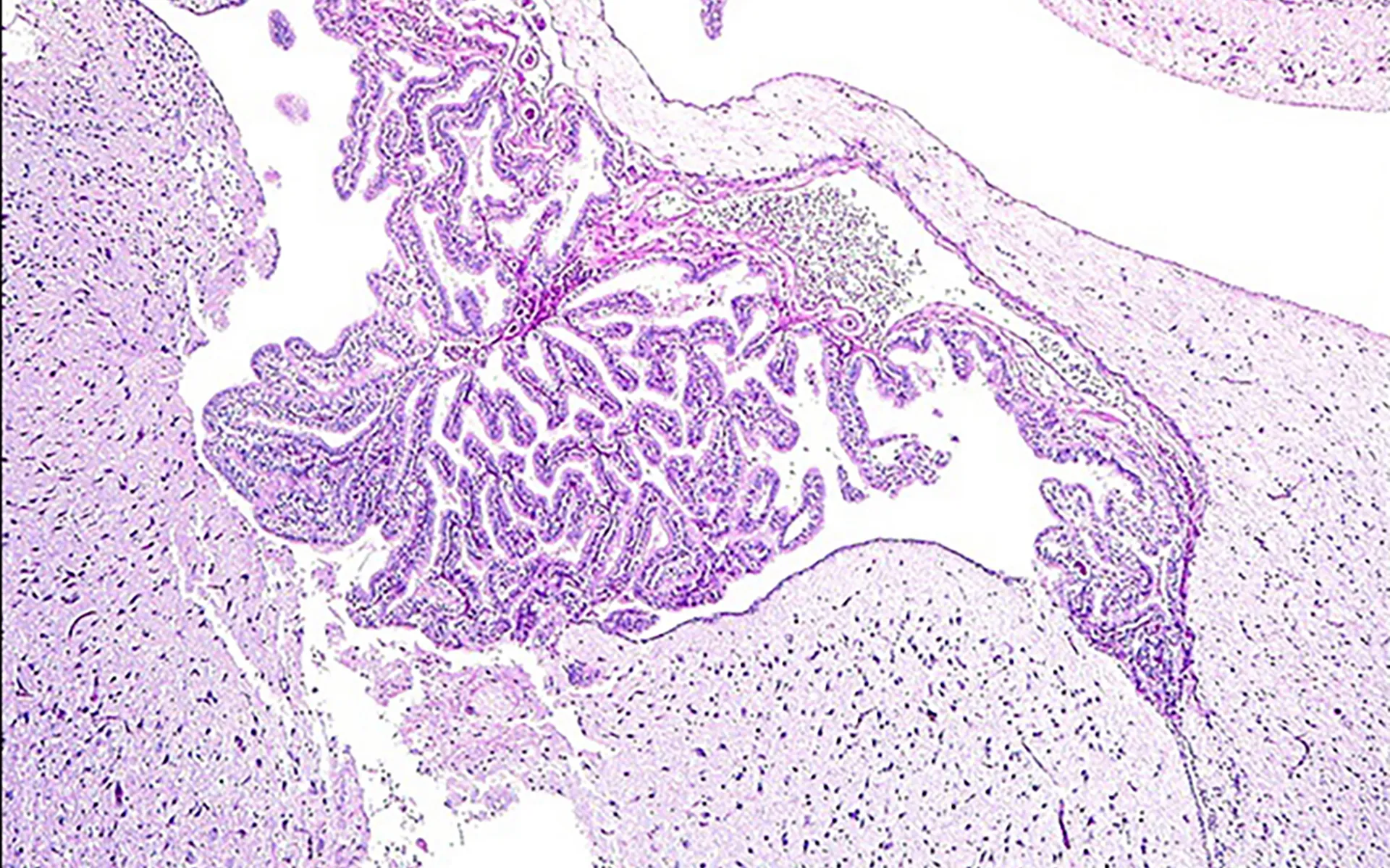
Choroid plexus tumors (CPT) are rare neoplasms and histologically classified as choroid plexus papilloma (CPP), atypical choroid plexus papilloma (APP), and choroid plexus carcinoma (CPC). These neoplasms have been described in humans, domestic animals (canine, feline, equine, caprine) and some wild animals (cetaceans and Psittacidae birds). To our best knowledge, herein we report the first CCP in a free-ranging eared dove (Columbiformes; Zenaida auriculata, de Murs 1847). Histologically, the ventricle was dilated, with a papillary proliferation (arboriform pattern) in topography of CP. The neoplasm was well-differentiated, composed by a single layer of cuboidal cells, anchored in a delicate fibrovascular stroma. The neoplastic cells exhibited moderate stroma, with well-defined borders and round nuclei, with vesicular chromatin and inconspicuous nucleoli. Mitotic activity was low (<1 mitosis per 10 high-power fields). Immunohistochemistry for cytokeratin markers (AE1/ AE3 antibody) were implemented, however, both neoplastic cells and normal epithelial tissues do not show immunoreactivity.

Downloads

Published

2021-11-30

Issue

Section

Artigos

How to Cite

Navas-Suárez, P. E., Ervedosa, T. B., di Loreto, C., Ressio, R. A., Jesus, I. P. de, Silva, M. M. H. da, Frediani, M. H., Guerra, J. M., & Fernandes, N. C. de A. (2021). Choroid plexus papilloma in a free ranging eared dove (Zenaida auriculata). Brazilian Journal of Veterinary Pathology, 14(3), 188-190. https://doi.org/10.24070/bjvp.1983-0246.v14i3p188-190