Diffuse melanosis secondary to metastatic melanoma in a Nelore bull

Authors

  • Jacqueline de Jesus Faculdade de Medicina Veterinária da Universidade Federal da Fronteira Sul (UFFS), Realeza, PR Author
  • Andrieli Bortolini Faculdade de Medicina Veterinária da Universidade Federal da Fronteira Sul (UFFS), Realeza, PR Author
  • Gabriela Corrêa de Almeida Faculdade de Medicina Veterinária da Universidade Federal da Fronteira Sul (UFFS), Realeza, PR Author
  • Bárbara Cardoso de Oliveira Faculdade de Medicina Veterinária da Universidade Federal da Fronteira Sul (UFFS), Realeza, PR Author
  • Artur Bruzamarelo Médico Veterinário autônomo Author
  • Raquel Rubia Rech Department of Veterinary Pathobiology, College of Veterinary Medicine & Biomedical Sciences, Texas A&M University, College Station, TX 77843-4467, USA Author
  • Claudio Severo Lombardo de Barros Faculdade de Medicina Veterinária e Zootecnia, Universidade Federal de Mato Grosso do Sul (UFMS), Campo Grande, MS. 5 Instituto de Ciências da Saúde, Universidade Paulista (UNIP), Bauru, SP Author
  • Carlos Eduardo Fonseca Alves Instituto de Ciências da Saúde, Universidade Paulista (UNIP), Bauru, SP Author
  • Fabiana Elias Faculdade de Medicina Veterinária da Universidade Federal da Fronteira Sul (UFFS), Realeza, PR Author

DOI:

https://doi.org/10.24070/bjvp.1983-0246.v14i3p191-198

Keywords:

Malignant melanoma, diffuse melanosis, immunohistochemistry, diseases of cattle

Abstract

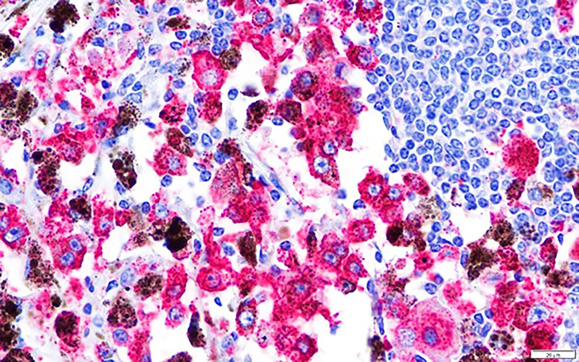
We report a case of diffuse melanosis secondary to metastatic malignant melanoma in a Nelore bull. Clinical signs included isolation from the herd, epistaxis, hyperthermia, pale ocular membranes, mucoid diarrhea and dark urine. Despite anti-inflammatory and antibiotic therapy, the bull died 45 days after the onset of the clinical signs. The most striking lesion was diffuse black discoloration to the visceral organs including liver, spleen, lungs, lymph nodes, and kidneys; all these affect organs were moderately enlarged”. The urine was black. Histologically, 50-80% of the parenchyma of the liver, spleen and lymph nodes was obliterated by aggregates of melanin-loaded neoplastic melanocytes. Those neoplastic cells also occurred within capillaries of the liver, spleen, lymph nodes, urinary bladder, lungs and kidneys. Immunohistochemistry of neoplastic melanocytes was positive for Melan A and PNL2 markers. Abundant brown to black pigment was found in melanophages in the lungs, confirmed by IBA1 immunohistochemistry.

Downloads

Published

2021-11-30

Issue

Section

Artigos

How to Cite

Jesus, J. de, Bortolini, A., Almeida, G. C. de, Oliveira, B. C. de, Bruzamarelo, A., Rech, R. R., Barros, C. S. L. de, Alves, C. E. F., & Elias, F. (2021). Diffuse melanosis secondary to metastatic melanoma in a Nelore bull. Brazilian Journal of Veterinary Pathology, 14(3), 191-198. https://doi.org/10.24070/bjvp.1983-0246.v14i3p191-198