Non jaagsiekte retrovirus associated adenosquamous lung carcinoma in a goat
DOI:
https://doi.org/10.24070/bjvp.1983-0246.v15i1p44-49Keywords:
retrovirus, adenomatous, squamous, lung neoplasm, small ruminantAbstract
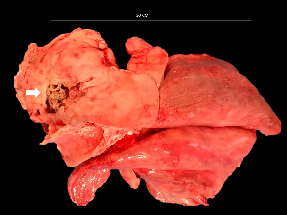
An 11-year-old pet goat presented a clinical history of acute respiratory distress with ultrasound diagnosis of wide lung injury. The animal was euthanized due to welfare reasons. At necropsy, it was found pleural effusion and adhesion on the right cranioventral thoracic region. The right cranial and middle lung lobes were firm and light gray with a neocavity containing purulent exudate. From the middle lobe, there was a nodular proliferation occupying alveolar spaces, densely cellular and composed by cuboid-columnar epithelial cells arranged in papillae and acini (60%), as well as polygonal cells arranged in nests with squamous differentiation (40%). Marked pleomorphism, anisocytosis and anisocariosis were also noted. A total of 39 mitosis figures for ten fields at 400x magnification were counted. Lung samples were negative for jaagsiekte retrovirus (JSRV) by PCR. Immunostaining for TTF1 and P53 occurred in zones of adenomatous and squamous differentiation, respectively. In MIB-1, 14% (82/594) of immunolabeled cells were observed in the squamous component. In conclusion, the histopathological and immunohistochemical characteristics confirmed the diagnosis of a pulmonary adenosquamous carcinoma, without JSRV involvement, in goat species.