Primary Tracheal Squamous Cells Carcinoma in a Dog
DOI:
https://doi.org/10.24070/bjvp.1983-0246.v15i3p147-152Keywords:
neoplasm, canine, histopathologyAbstract
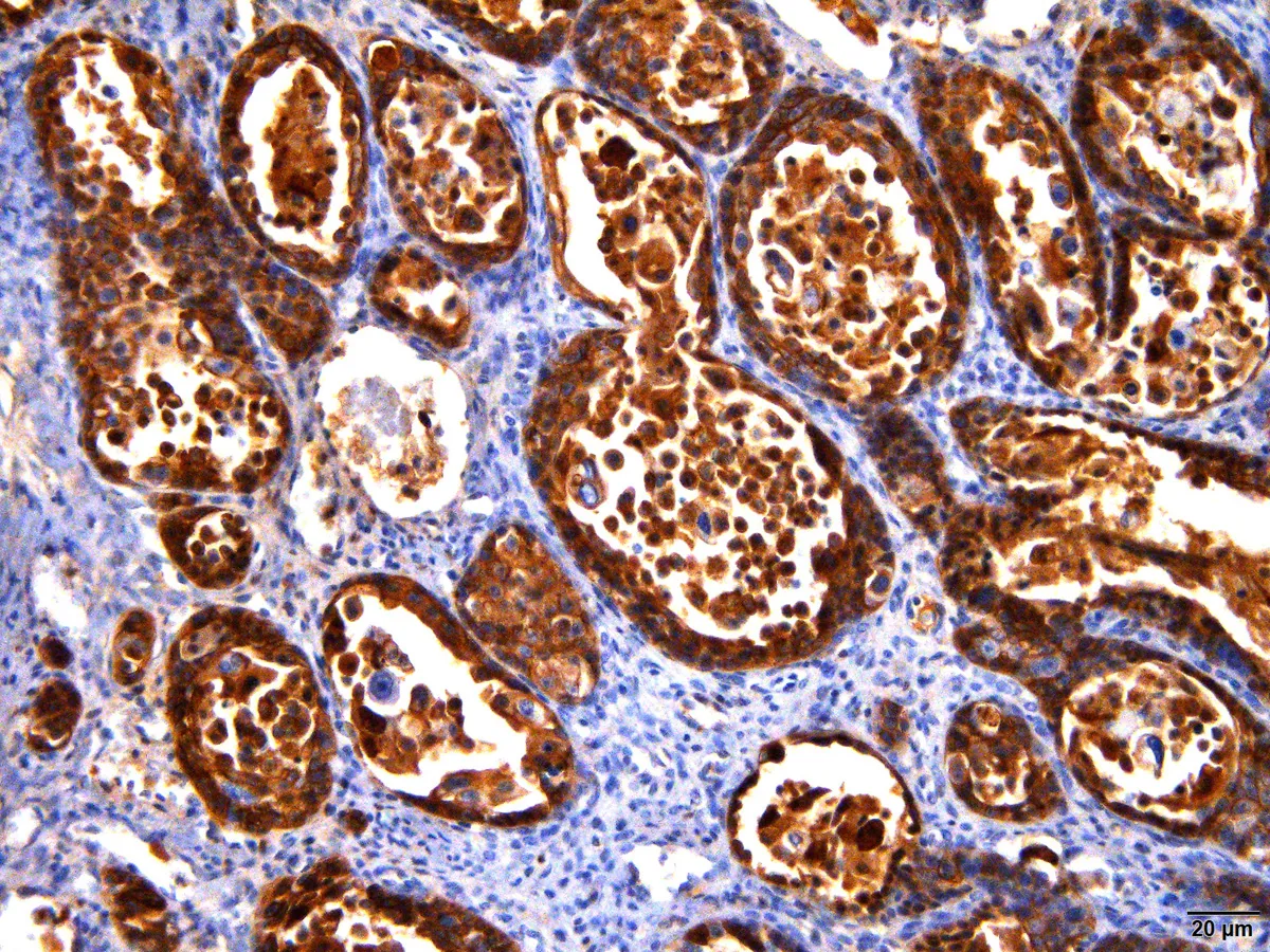
Primary tracheal neoplasms are uncommon and poorly documented in veterinary medicine, being most frequently reported in domestic cats. Squamous cell carcinoma (SCC) is a malignant neoplasm that originates from the stratified squamous epithelium, considered one of the most common skin neoplasms in dogs and cats. This paper aims to report the anatomopathological and immunohistochemical findings of a clinical case of primary SCC in the trachea of a female Schnauzer canine, attended at the Veterinary Hospital Luiz Quintiliano de Oliveira of the Faculty of Veterinary Medicine of Araçatuba (FMVA – UNESP), presenting clinical signs such as choking, coughing and dyspnea for 1 month. The tracheal portion affected by the neoplasm was sent to the Veterinary Pathology department of the FMVA after surgical excision. Fragments of the neoplasm were collected and fixed in 10% formaldehyde for further histopathological and immunohistochemical analysis. Microscopically, the neoplasm was well differentiated, being characterized by the presence of keratin pearls, low degree of pleomorphism and rare mitotic figures. In the immunohistochemical analysis, there was immunoexpression of anti-cytokeratin antibodies AE1AE3, 34BE12, CK14 and CK5/6, confirming the diagnosis of squamous cell carcinoma. In about 30% of the cells there was immunostaining for Ki67 antibodies, justifying the low mitotic index of tumor cells and the few images of mitosis seen. Due to the rare occurrence of primary tracheal SCC in dogs, the use of diversified diagnostic techniques is important in order to better understand the biological behavior of this neoplasm in unusual anatomical locations.