
Histopathological case study of canine hemangiosarcoma with multiple organ metastases
DOI:
https://doi.org/10.24070/bjvp.1983-0246.v15i3p168-172Keywords:
dog, pathology, hemangiosarcoma, spleenAbstract
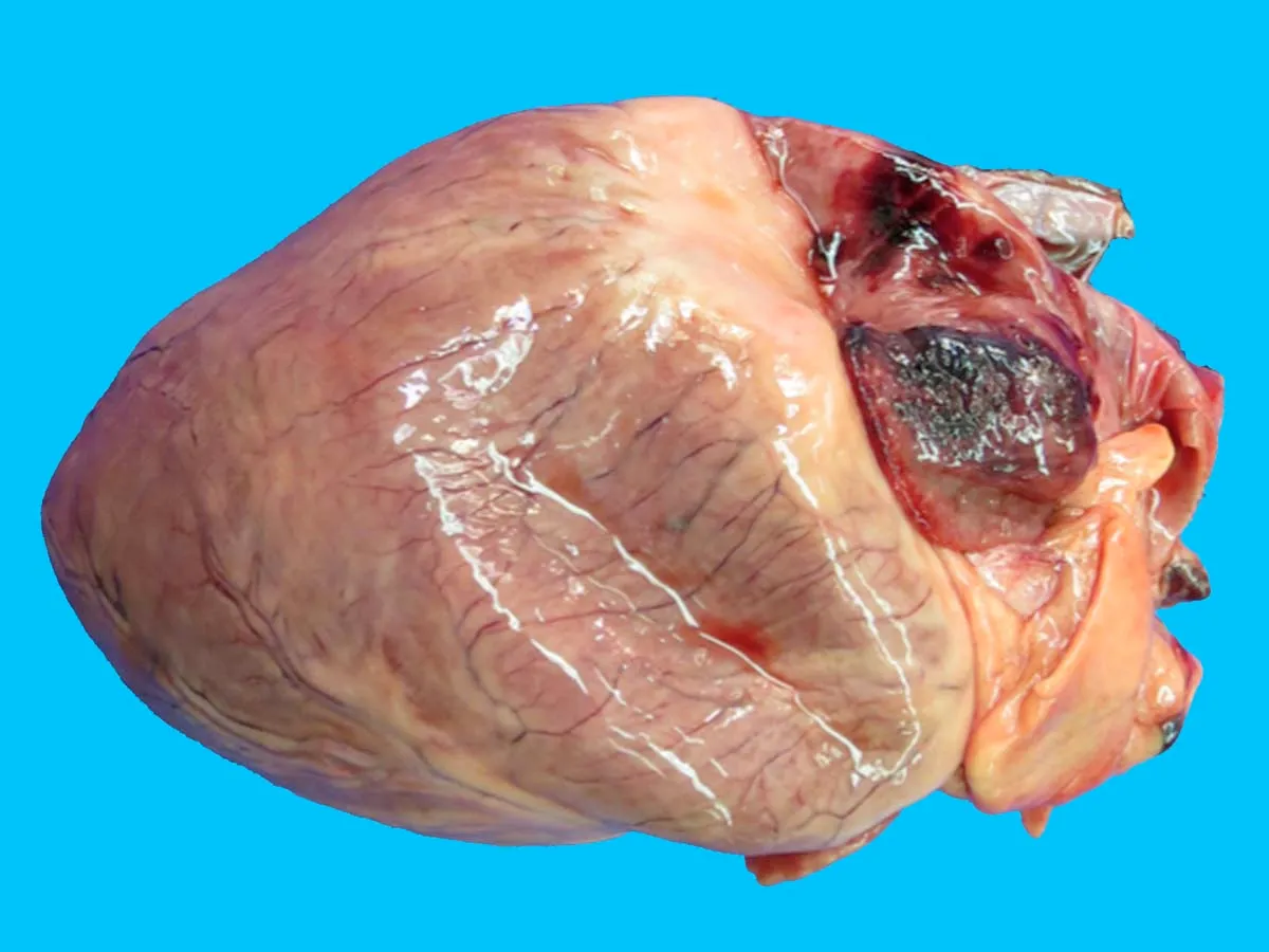
We studied a 14-year-old male dog necropsied. Gross findings were hemorrhagic nodules in the spleen, liver, heart and abdominal and thoracic lymph nodes. Histologically, we homogeneously observed tumor cells often with prominent, bulging and mitotic nuclei that were pleomorphic and hyperchromatic, forming small blood vessels. Tumors in all organs were diagnosed as capillary hemangiosarcomas. No tumor cells were detected in the lungs. We presume that the primary tumor was present in the spleen, from where it metastasized multiple organs via lymphatic vessels.
Downloads
Published
2022-11-30
Issue
Section
Articles
How to Cite
Varela, B., Larrañaga, C., Yamasaki, K., & Verdes, J. M. (2022). Histopathological case study of canine hemangiosarcoma with multiple organ metastases. Brazilian Journal of Veterinary Pathology, 15(3), 168-172. https://doi.org/10.24070/bjvp.1983-0246.v15i3p168-172