Age, and non-age-related cytoarchitectural changes in the brain of some Nigerian cattle
DOI:
https://doi.org/10.24070/bjvp.1983-0246.v16i1p35-45Keywords:
age-related, brain, cattle, changes, NigeriaAbstract
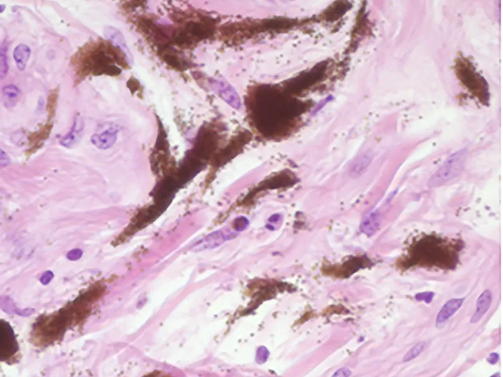
Brain is the most vulnerable organ in the body to the ageing processes which operates at variable levels between organs and species. In this study we examine brains histopathologically for architectural changes in four Nigerian cattle breeds. Brains from 246 cattle (Bunaji=80, Muturu=78, Rahaji=62 and Sokoto Gudali=26), aged 1–≥12years were examined at 8 different neuroanatomical locations. All the cattle used were obtained from Abattoir in Ibadan, Nigeria. They were examined at ante-mortem and those without clinical signs suggestive of neurological disease were sampled. Major changes observed were intracellular accumulation of substances (61.4%), neuronal and or axonal degenerations and loss (51.6%), perivascular cuffing (50.0%), extracellular accumulation of substances (29.7%), hypercellularity (19.1%) and spongy state (0.4%), malacia (0.0%), demyelination (0.0%) in 233 cattle. Intraneuronal vacuolations (35%), lipofuscin accumulation (42%), axonal spheroids (50%), inflammatory changes (50%), and brain sand (22%). Although perivascular cuffing was high, there was low incidence of lymphocytic infiltration in the pineal gland in both sexes. Intracellular accumulation of substances, neuronal and or axonal degenerations and loss, and extracellular accumulation of substances display levels of significant changes with age (p<0.0001). Changes starts at the age of 2 years in life in these breeds of cattle due probably to the adverse stress exerted by the tropical climate. The description of histological findings in the brain of symptomless cattle in the present study provides a useful background for diagnostic bovine neuropathology in the tropics.