Comparative study of parasite load in the spleen, lymph node, and skin of dogs with visceral leishmaniasis
DOI:
https://doi.org/10.24070/bjvp.1983-0246.v17i2p84-92Keywords:
zoonosis, immunohistochemistry, histopathology, Leishmania sp., immune response, correlation, dogAbstract
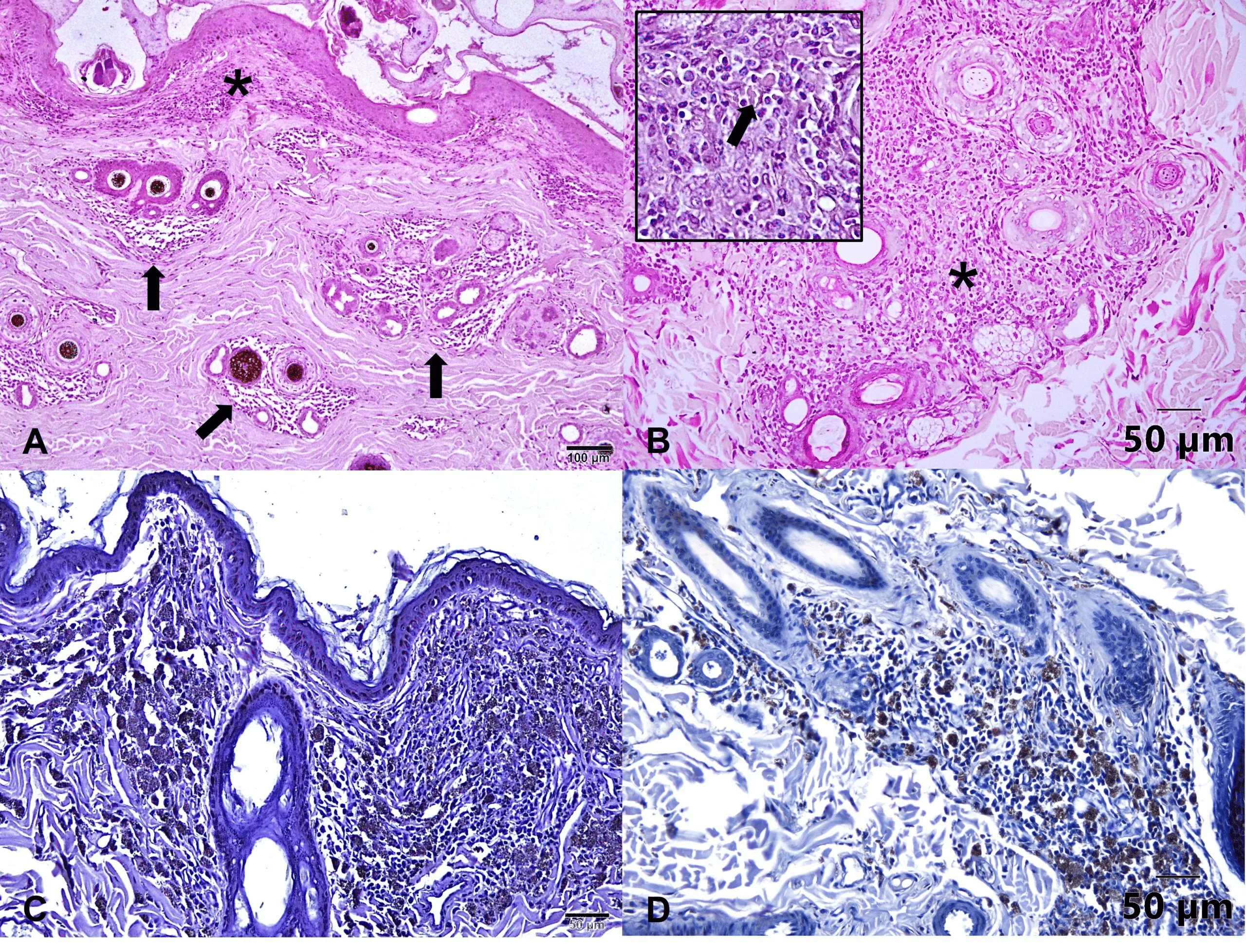
Canine visceral leishmaniasis (VL) is a zoonosis caused by the protozoan Leishmania infantum. The lymph nodes, spleen, and skin are essential organs in the immunopathogenesis of the disease. This study aimed to investigate the histomorphological alterations and parasite load in the popliteal lymph node, spleen, and skin of eleven VL-positive dogs in the fine needle aspiration (FNA), Dual-path Platform chromatographic immunoassay (DPP® CVL rapid test) and Enzyme-linked immunosorbent assay (ELISA). Histopathological and immunohistochemical methods were used to evaluate the samples, and the results showed variable histopathological changes and parasite load. The popliteal lymph nodes and spleen exhibited granulomatous reaction, lymphoid atrophy, presence of plasma cells, and disorganization of the architecture was marked. The skin showed multifocal to diffuse inflammation in the superficial dermis, composed of lymphoplasmacytic infiltrate and granulomatous reaction. Immunodetection of the parasite Leishmania sp. was observed in all organs. The intensity of histological changes was not associated with the higher number of parasitized macrophages. The popliteal lymph node had the highest median parasite load (11.2) compared to the skin and spleen. Statistically, the Pearson correlation test revealed a highly significant correlation in the parasite load between the popliteal lymph node and spleen (r=0.89081, p=0.0002) and between the popliteal lymph node and skin (r=0.71185, p=0.0140). The study concludes that VL-positive dogs’ lymph nodes, spleen, and skin suffer histomorphological alterations that could be one of the aspects that favor the maintenance of the infection.


