Histopathological lesions in Corydoras spp. (Siluriformes: Callychthyidae) caused by Procamallanus (Spirocamallanus) pintoi (Nematoda: Camallanidae) from Iquitos, Peru
DOI:
https://doi.org/10.24070/bjvp.1983-0246.v17i3p179-188Keywords:
Aquaculture, Amazon, histopathological lesions, necrosis, fish healthAbstract
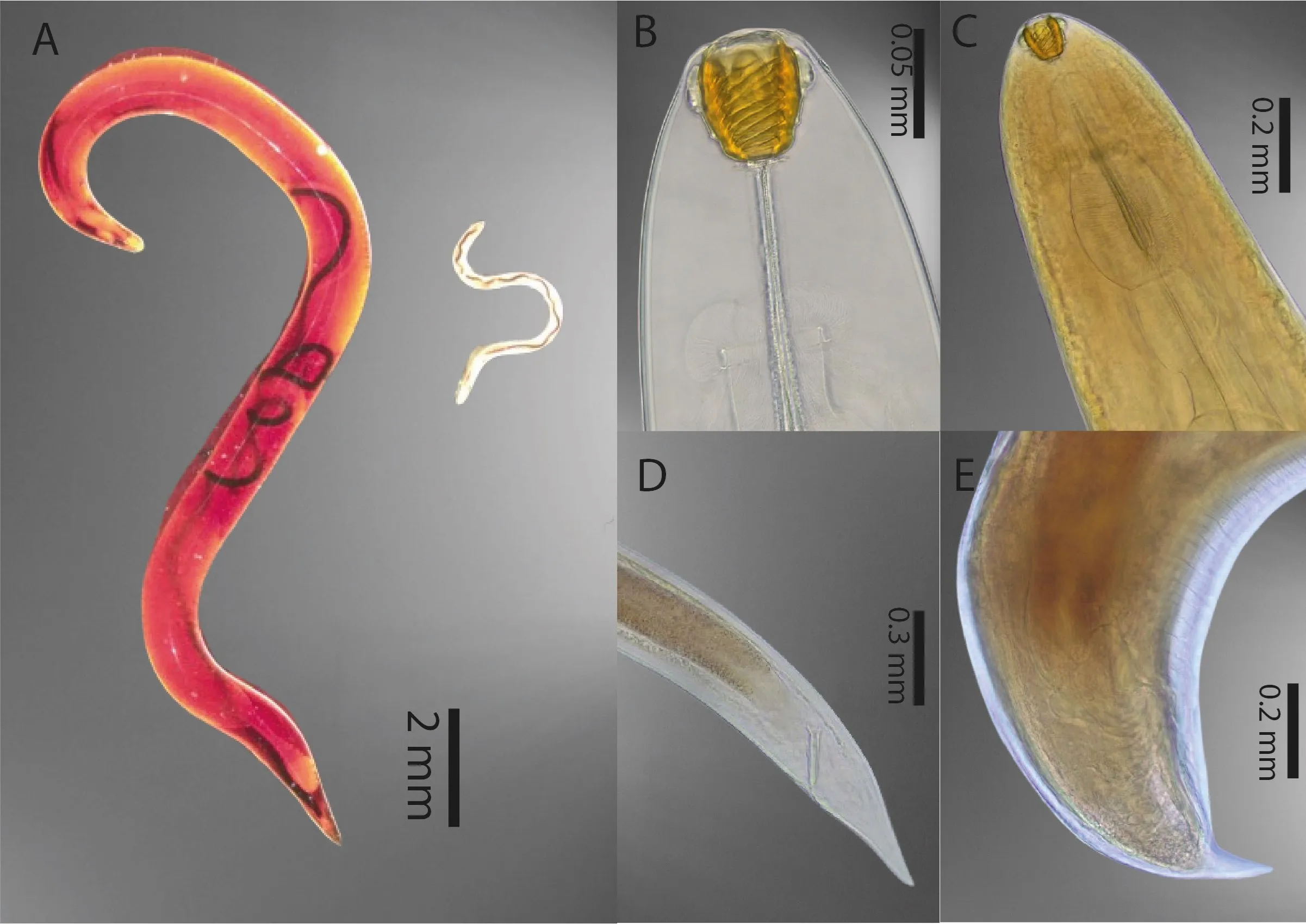
Callichthyidae is a family of armored catfishes commercially important in the Peruvian Amazon. Among these species, Corydoras acutus, C. reticulatus, and C. virginiae are stored for weeks or months and then exported to different countries worldwide, where they may present some sanitary problems along the way, such as parasitic infections. In the present study, it was reported the level of infection of Procamallanus (Spirocamallanus) pintoi (Kohn and Fernandes, 1988), through the calculation of their parasitological indices and the description of their histopathological lesions in the intestinal tract. Sixty individuals of each species were acquired from fishermen of the District of Belén, in Iquitos, Peru. Fish were transported to the “Laboratorio de Parasitología y Sanidad Acuícola” from the “Instituto de Investigaciones de la Amazonía Peruana” (IIAP), in Iquitos-Peru. Parasitological examination allowed us to identify the nematode P. pintoi infecting the intestinal tract of the fish. Histopathological analysis revealed: inflammatory cell infiltration (73.3%, 63.3%, and 60%), enterocyte hyperplasia (90%, 70%, and 63.3%), desquamation of epithelial cells (90%, 70%, and 63.3%), goblet cell hyperplasia (76.7%, 66.7%, and 50%) and necrosis (70%, 60%, and 66.7%), in C. acutus, C. reticulatus, and C. virginiae respectively for each lesion.