Rhinosporidium seeberi infection in a male mule in the State of Tocantins (Brazil)

Authors

  • Taismara Simas de Oliveira Laboratório Hermes Pardini, Belo Horizonte Author
  • Silvia de Araújo França Baêta Departamento de Medicina Veterinária, Faculdade de Medicina, Universidade Federal de Juiz de Fora, Juiz de Fora Author
  • Rodolfo Olinto Rotoli Garcia de Oliveira Veterinary Practitioners, Araguaína Author
  • Antônio Araújo Leite Veterinary Practitioners, Araguaína Author
  • Cristiane América de Carvalho Animale Laboratório Veterinário, Araguaína Author
  • Jefferson Bruno Soares Oliveira Departamento de Clínica e Cirurgia Veterinárias, Escola de Veterinária, Universidade Federal de Minas Gerais, Belo Horizonte Author
  • Tatiane Alves da Paixão Departamento de Patologia Geral, Instituto de Ciências Biológicas, Universidade Federal de Minas Gerais, Belo Horizonte Author
  • Renato Lima Santos Departamento de Clínica e Cirurgia Veterinárias, Escola de Veterinária, Universidade Federal de Minas Gerais, Belo Horizonte Author

DOI:

https://doi.org/10.24070/bjvp.1983-0246.018003

Keywords:

rhinosporidiosis, Mesomycetozoan, nasal polyps, rhinitis, granulomatous

Abstract

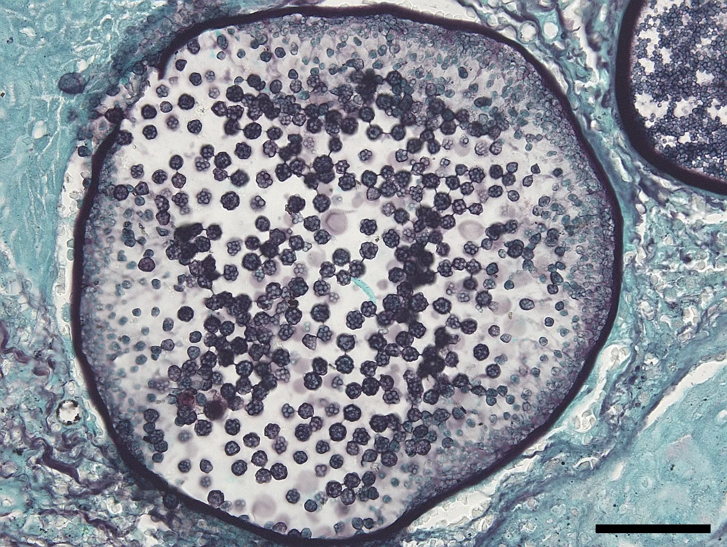
Rhinosporidiosis is a chronic disease caused by Rhinosporidium seeberi, which can infect several host species, including domestic animals and humans. The taxonomy of R. seeberi is still controversial, but scientific evidence supports the classification of this organism as a Mesomycetozoan. Considering the absence of previously reported cases of rhinosporidiosis in the Northern Region of Brazil, the goal of this report was to describe the clinical and pathological findings of a case of R. seeberi infection in a male mule in Araguaína (Tocantins State, Brazil). An 8-year-old male mule presented respiratory difficulties due to a polypoid exophytic lesion that partially obstructed the nasal cavity. The lesion was surgically excised and submitted for microscopic analysis. Microscopically, there was multifocal erosion, epithelial hyperplasia, diffuse inflammatory infiltrate, composed of many histiocytes and plasma cells, and fewer neutrophils and lymphocytes, with numerous intralesional organisms with diameters ranging from 10 to 400 μm compatible with sporangia exhibiting numerous nuclei and endoconidia compatible with R. seeberi.

Downloads

Published

2025-03-30

Issue

Section

Artigos

How to Cite

Oliveira, T. S. de, Baêta, S. de A. F., Oliveira, R. O. R. G. de, Leite, A. A., Carvalho, C. A. de, Oliveira, J. B. S., Paixão, T. A. da, & Santos, R. L. (2025). Rhinosporidium seeberi infection in a male mule in the State of Tocantins (Brazil). Brazilian Journal of Veterinary Pathology, 18, e018003. https://doi.org/10.24070/bjvp.1983-0246.018003