
Prolonged survival in a dog with unresectable exocrine pancreatic adenocarcinoma treated with toceranib phosphate: a case report
DOI:
https://doi.org/10.24070/bjvp.1983-0246.019011Keywords:
Pancreatic adenocarcinoma, toceranib phosphate, multikinase panel, precision medicineAbstract
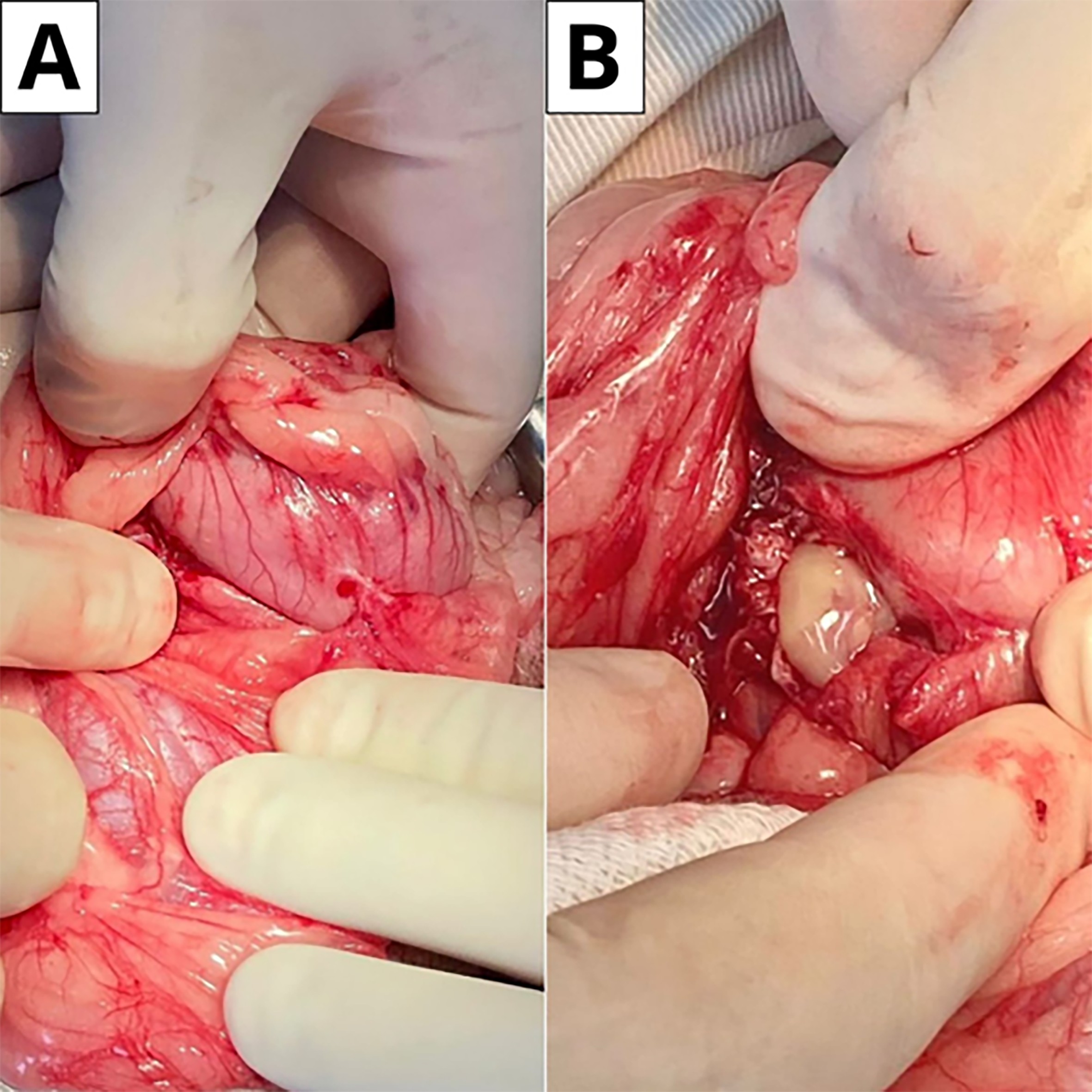
Canine pancreatic carcinoma is an uncommon and highly aggressive tumor usually detected at an advanced stage. This case report describes a dog with exocrine pancreatic adenocarcinoma that presented with diarrhea, vomiting, abdominal pain, and anorexia. Computed tomography (CT) revealed pancreatic enlargement with nodular formation in the body and left lobe of the pancreas. Resection was not feasible because of the tumor location, and incisional biopsy was performed. Histopathology demonstrated large polygonal neoplastic cells arranged in a disorganized manner, forming clusters and acinar structures, consistent with exocrine pancreatic adenocarcinoma. As no effective medical treatment exists for this condition, a multikinase immunohistochemical panel was used to guide therapy. The panel revealed overexpression of the vascular endothelial growth factor receptor (VEGFR, Score 4+) and activation of the mitogen-activated protein kinase (MAPK/Erk1/2) pathway (score 3+). Based on these findings, toceranib phosphate was initiated at 2.75 mg on a Monday, Wednesday, Friday (MWF) schedule. This targeted therapy resulted in a partial response on ultrasound, with the pancreatic lesion decreasing from 2.63 × 2.89 cm to 1.75 × 1.56 cm after 66 days and further reducing to 1.11 × 1.40 cm at 122 days. From day 213 onward, the lesion was no longer detected on the follow-up ultrasound. However, complete remission cannot be confirmed without histopathological reassessment or advanced imaging such as computed tomography. The patient remains alive with a survival time of 484 days under ongoing monitoring. Despite this encouraging outcome, further studies are required to evaluate the efficacy of tyrosine kinase inhibitors in managing canine exocrine pancreatic adenocarcinoma.
Downloads
Published
Issue
Section
License
Copyright (c) 2026 Brazilian Journal of Veterinary Pathology

This work is licensed under a Creative Commons Attribution 4.0 International License.