
Canine myxoid mesothelioma with clinical presentation of Pseudomixoma peritonei
DOI:
https://doi.org/10.24070/bjvp.1983-0246.018014Keywords:
gelatinous extracellular matrix, mesenchymal neoplasms, mesothelial, myxoidAbstract
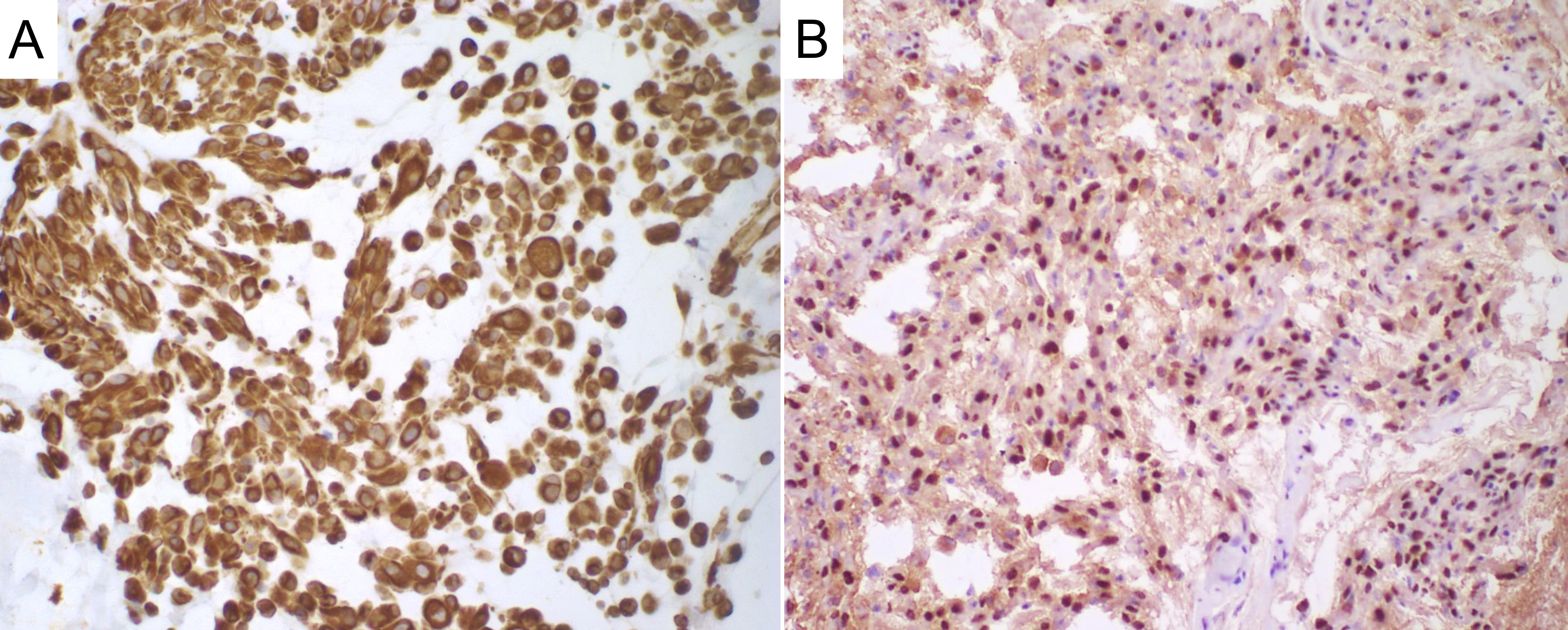
Myxoid mesothelioma is a rare variant of mesothelioma, characterized by a myxoid component, which refers to a gelatinous extracellular matrix containing mucin. This study describes a case of myxoid mesothelioma in a 13-year-old Yorkshire Terrier that presented with sudden abdominal distension, pain, and respiratory discomfort. Imaging exams revealed an extensive amount of amorphous, homogeneous, and viscous material in the abdominal cavity. Due to unsuccessful drainage attempts, exploratory laparoscopy was performed, uncovering large volumes of gelatinous material and areas of mesenteric thickness. Despite intensive care, the animal died post-surgery and was submitted for necropsy. Gross examination revealed diffuse gelatinous material coating the omentum and mesentery, along with firm micronodular areas. Microscopic analysis showed neoplastic sarcomatoid to round cells supported by delicate fibrovascular connective tissue with abundant myxoid stroma. Marked anisocytosis, anisokaryosis and cellular pleomorphism were observed; the myxoid nature of the stroma was confirmed by intense Alcian blue staining. Immunohistochemistry revealed positive vimentin and WT1 immunoreactivity, confirming the mesothelial origin of the tumor, while pancytokeratin, carcioembryonic antigen, high molecular weight cytoqueratin, and calretinin were negative. These findings support the diagnosis of myxoid-variant mesothelioma with clinical presentation of Pseudomyxoma peritonei.
Downloads
Published
Issue
Section
License
Copyright (c) 2025 Brazilian Journal of Veterinary Pathology

This work is licensed under a Creative Commons Attribution 4.0 International License.