
Establishing primary tumor cell lines in dogs: Challenges and Ki-67 correlation
DOI:
https://doi.org/10.24070/bjvp.1983-0246.019004Keywords:
cancer, molecular markers, lineages, translational researchAbstract
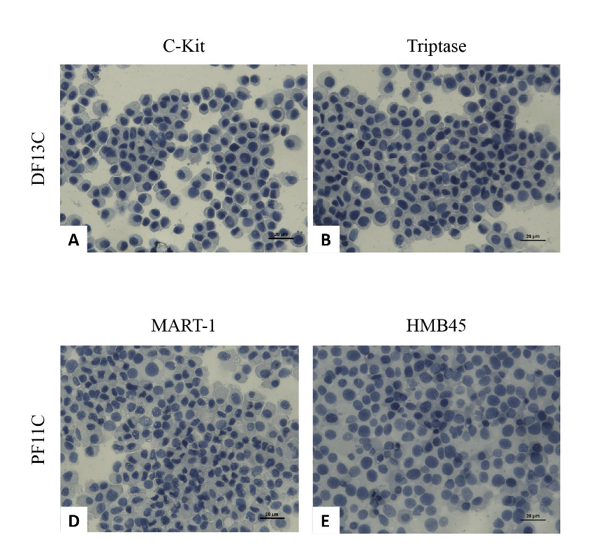
The incidence of malignant neoplasms in dogs has significantly increased in recent decades. Due to their aggressive nature and poor prognosis, the search for new, patient-specific treatments has become a promising approach. The use of a patient’s own tumor cell lines is a valuable tool for genetic analysis, treatment targeting, and serves as a translational strategy for the development of new drugs. However, there are technical limitations and poorly understood factors that influence the success or failure of establishing cell lines from primary tumors. This study aimed to establish continuous tumor cell lines from canine patients using a simplified protocol. After 10 passages in culture, the cells were considered established and used for further assays. Of the 10 tumors collected, 6 cell lines were successfully established: oral melanoma (PM13C and PF11C), cutaneous lymphoma (BF11C), mast cell (DF13C), urothelial carcinoma (MF10C), and mammary carcinoma (PF12C). Statistical analysis using the Chi-squared test revealed that success in establishing the cell lines was correlated with the histopathological grade of malignancy and the clinical stage of the patients, but not with the Ki-67 proliferation marker. DF13C and PF11C cells were inoculated into a xenograft model and exhibited a neoplastic pattern, confirming the histogenesis of the lines. This study identifies key factors influencing the success or failure of establishing long-term cell lines from patient tumors, contributing valuable data that is currently scarce in the literature and limiting the integration of this protocol into routine laboratory practices.
Additional Files
Published
Issue
Section
License
Copyright (c) 2026 Brazilian Journal of Veterinary Pathology

This work is licensed under a Creative Commons Attribution 4.0 International License.