Bovine Abortion Associated with Neospora caninum
Diagnosis and Epidemiological Aspects of a Dairy Cattle Herd in the Northeast Region of São Paulo State, Brazil
DOI:
https://doi.org/10.24070/bjvp.1983-0246.004018Keywords:
Neosporosis, cattle, abortionAbstract
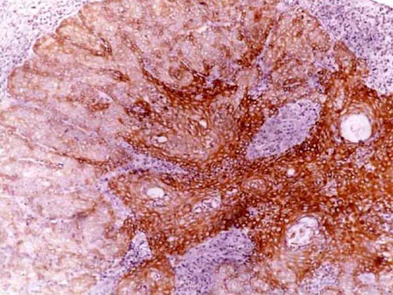
The objective of this study was to report the presence of Neospora caninum-associated abortion in bovines at a farm in the northeast region of São Paulo State. In January 2010, it was sent to the Department of Pathology, UNESP-Jaboticabal, a bovine fetus with an estimated age of seven months, which was natural of a dairy farm with 300 animals and an average daily production of 3,000 liters of milk, nearly 20 liters per cow. The animals were vaccinated against rabies, foot and mouth disease, carbuncle, brucellosis, leptospirosis, bovine herpes virus type I and bovine viral diarrhea virus. The herd consisted of purebred Holstein animals, Jersey, and mostly by crossbred animals 7-8 (gir x holstein). During necropsy, samples of the serosanguineous liquid present at the thoracic cavity and the heart of the fetus were collected for the detection of anti-Neospora caninum antibodies through Indirect Immunofluorescence Assay (IFA). Fragments of brain, cerebellum, tongue, liver, heart and kidneys were collected for the execution of histopathology (HP), immunohistochemistry (IHC) and Polymerase Chain Reaction. In order that IFA could be performed, the owner was requested blood samples without anticoagulants of the mother and other cows in the farm, with or without a history of abortion. At necropsy, it was verified a severe autolysis of the fetus. The serology of the fetus was 1:25, while the serology of the mother was 1:3,200. At HP, it was observed discrete multifocal non-suppurative encephalomyelitis characterized by gliosis and mononuclear inflammatory infiltration associated with cellular debris. DNA amplification of N. caninum was positive in fragments of brain, tongue, cerebellum, heart and kidneys. At IHC, it has been observed immunoreactivity to a cyst located in the tongue. The owner reported that his herd showed endemic episodes of abortion, while 27.69% (18/65) of the 65 animals sampled were seropositive. Although it has not been a significant difference (p>0.05), a higher seropositivity was observed in animals with a history of abortion (10/26) 38.46%, in comparison with animals without previous abortion (8/39) 20.51%. These findings show that the abortion under study was provoked by the protozoan N. caninum, while this is the first report concerning cattle in the northeast region of São Paulo State.