Mixed Thymoma in a Goat

Authors

  • Selwyn A. Headley Section of Veterinary Pathology, Department of Basic Veterinary Sciences, University of Helsinki, Finland Author
  • Antti Sukura Section of Veterinary Pathology, Department of Basic Veterinary Sciences, University of Helsinki, Finland Author

Keywords:

goat, thymoma, immunohistochemistry, pathology

Abstract

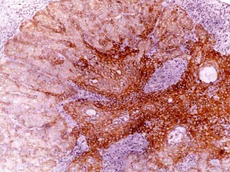
This article describes the gross, histopathological, and immunohistochemical findings of a mixed thymoma in a 2-year-old, mixed-breed, female goat. A large space-occupying neoplastic growth was observed within the cranial thoracic cavity, which by histology was well-encapsulated and formed by the proliferation of spindle-shaped epithelial cells arranged in solid sheets or rosette-like formations with varying accumulations of lymphocytes. Immunohistochemistry demonstrated that the neoplastic epithelial cells expressed cytokeratin, with negative immunoreactivity to vimentin, thyroglobulin, Chromogranin A, neuron-specific enolase, and glial fibrillary acidic protein. The lymphocytic population expressed CD3 and CD2. These findings favor a diagnosis of mixed thymoma.

Author Biographies

  • Selwyn A. Headley, Section of Veterinary Pathology, Department of Basic Veterinary Sciences, University of Helsinki, Finland

    Section of Veterinary Pathology, Department of Basic Veterinary Sciences, University of Helsinki, Finland

  • Antti Sukura, Section of Veterinary Pathology, Department of Basic Veterinary Sciences, University of Helsinki, Finland

    Section of Veterinary Pathology, Department of Basic Veterinary Sciences, University of Helsinki, Finland

Downloads

Published

2011-07-30

Issue

Section

Artigos

How to Cite

Headley, S. A., & Sukura, A. (2011). Mixed Thymoma in a Goat. Brazilian Journal of Veterinary Pathology, 4(2), 126-131. https://bjvp.org.br/bjvp/article/view/89