Giant cutaneous horn associated with a dilated pore in a cat

Authors

  • Millena Ramos Alves Pathologic Anatomy Sector, Department of Epidemiology and Public Health, Institute of Veterinary, Federal Rural University of Rio de Janeiro Author https://orcid.org/0009-0002-8362-1453
  • Adriano de Sales Araujo Pathologic Anatomy Sector, Department of Epidemiology and Public Health, Institute of Veterinary, Federal Rural University of Rio de Janeiro Author https://orcid.org/0009-0003-9447-8282
  • Paula Dias Retamero Pathologic Anatomy Sector, Department of Epidemiology and Public Health, Institute of Veterinary, Federal Rural University of Rio de Janeiro Author https://orcid.org/0009-0009-5011-0164
  • Karla Geovanna Pereira Nascimento Pathologic Anatomy Sector, Department of Epidemiology and Public Health, Institute of Veterinary, Federal Rural University of Rio de Janeiro Author https://orcid.org/0000-0002-0684-4998
  • Áster Patricia Kerschr Bento Pathologic Anatomy Sector, Department of Epidemiology and Public Health, Institute of Veterinary, Federal Rural University of Rio de Janeiro Author https://orcid.org/0009-0009-0153-2328
  • Vivian de Assunção Nogueira Carvalho Pathologic Anatomy Sector, Department of Epidemiology and Public Health, Institute of Veterinary, Federal Rural University of Rio de Janeiro Author https://orcid.org/0000-0002-4034-9675
  • Ticiana do Nascimento França Pathologic Anatomy Sector, Department of Epidemiology and Public Health, Institute of Veterinary, Federal Rural University of Rio de Janeiro Author https://orcid.org/0000-0002-7365-2697

Keywords:

feline, face, keratinous growth, projection, skin

Abstract

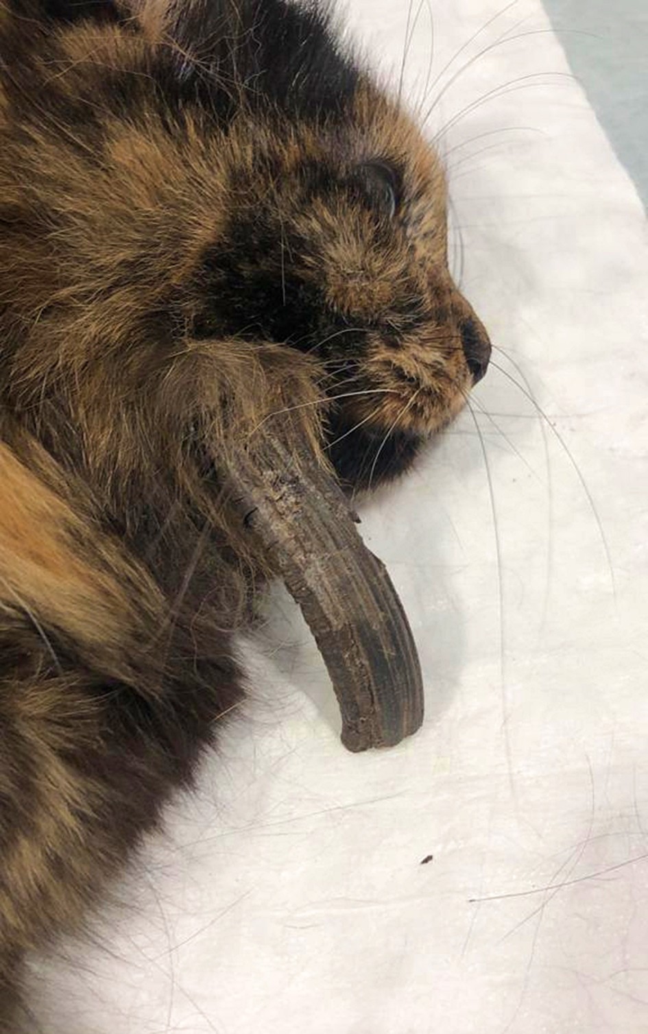
This report aims to describe a rare case of a giant cutaneous horn in a domestic cat and its association with a dilated pore. A 6-year-old female domestic cat was presented to a veterinary clinic with a keratinized, horn-like lesion on the right lateral cheek. The cylindrical mass measured 9.5 cm in length, 1.5 cm in width, and 1.5 cm in thickness, and appeared firm, dark brown to black, and hyperkeratotic. Histopathological examination revealed a dermal cyst lined by well-differentiated squamous epithelium with a prominent granular layer, consistent with an infundibular origin. The cystic lumen contained compact, laminated, partially pigmented keratin, with focal areas of parakeratosis. The lesion was characterized by keratinocyte hyperplasia with extensive laminated parakeratotic hyperkeratosis. These findings led to the diagnosis of a dilated pore, a proliferative variant of the feline infundibular follicular cyst, associated with a giant cutaneous horn. This rare presentation not only provides insight into the pathogenesis and potential relationship between dilated pores and cutaneous horns in cats, but also expands the current understanding of their differential diagnoses, particularly in atypical locations such as the face.

Published

2026-04-21

Issue

Section

Accepted manuscripts

How to Cite

Ramos Alves, M., de Sales Araujo, A., Dias Retamero, P., Geovanna Pereira Nascimento, K., Patricia Kerschr Bento, Áster, de Assunção Nogueira Carvalho, V., & do Nascimento França, T. (2026). Giant cutaneous horn associated with a dilated pore in a cat. Brazilian Journal of Veterinary Pathology, 19, e019015. https://bjvp.org.br/bjvp/article/view/533