
Transmural gastrointestinal stromal tumor of the mixed subtype in a canine: case report
Keywords:
dog, GIST, c-Kit, immunohistochemistry, mesenchymal neoplasmAbstract
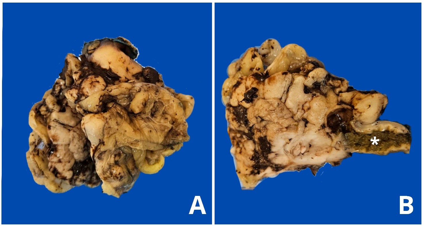
Gastrointestinal stromal tumors (GISTs) are mesenchymal neoplasms originating from interstitial cells of Cajal, characterized, in most cases, by the immunohistochemical expression of c-Kit (CD117). In dogs, they are rare and the associated clinical signs are nonspecific, including vomiting, diarrhea, weight loss, and abdominal pain, which often makes clinical diagnosis difficult. This paper describes a case of GIST in a dog, located in the cecum, detected through imaging exams and diagnosed via anatomopathological evaluation, and immunohistochemical. Macroscopically, the mass was transmural and measured approximately 7.0 cm in diameter, white to dark brown in color, had a soft consistency, and, upon cutting, a smooth, solid surface. Microscopically, the neoplasm was predominantly composed of spindle cells, with multifocal areas of epithelioid cells, both with positive plasma membrane staining for c-Kit and absence of Desmin expression, confirming the diagnosis. This report reinforces the importance of immunohistochemical evaluation in differentiating GISTs from other mesenchymal neoplasms of the gastrointestinal tract and contributes to the understanding of the morphological and clinicopathological aspects of this entity in dogs.
Downloads
Published
Issue
Section
License
Copyright (c) 2026 Brazilian Journal of Veterinary Pathology

This work is licensed under a Creative Commons Attribution 4.0 International License.