
Cervical stenotic myelopathy in a Brazilian Sport Horse foal
Keywords:
equine, Wobbler syndrome, computed tomographyAbstract
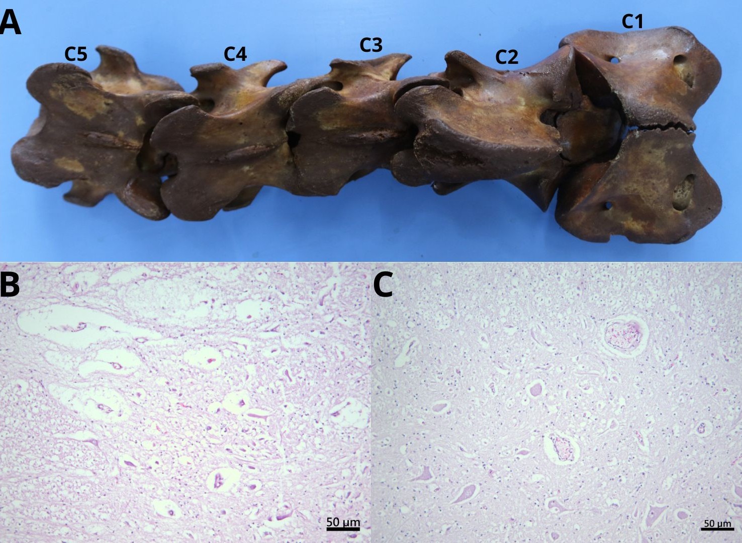
Cervical stenotic myelopathy (CSM) is a neurological disorder characterized by spinal cord compression in the cervical region, frequently associated with ataxia in horses. The objective of this study was to report a case of cervical stenotic myelopathy in a Brazilian Sport Horse filly evaluated at the Veterinary Hospital of the Federal University of Mato Grosso, Cuiabá campus, describing the clinical history, epidemiological factors, radiographic and tomographic findings of the cervical region, and post mortem findings. The animal presented with a history of frequent falls, incoordination and Increased volume in the femoral region of the right pelvic limb.. Laboratory tests showed no significant abnormalities, and swelling in the pelvic limb was compatible with a hematoma. Cervical radiographs revealed spinal canal stenosis between C3 and C4. After death, computed tomography associated with myelography demonstrated greater compression at this level during cervical flexion, with sagittal ratio values consistent with stenosis. Morphological evaluation of the cervical vertebrae indicated articular incongruence suggestive of cervical instability, and histopathology of the spinal cord revealed degenerative changes consistent with cervical stenotic myelopathy. These results highlight the importance of combining clinical evaluation, imaging studies, and anatomopathological analysis for definitive diagnosis and emphasize the diagnostic complexity of this condition, especially in young animals with nonspecific neurological signs.
Published
Issue
Section
License
Copyright (c) 2026 Brazilian Journal of Veterinary Pathology

This work is licensed under a Creative Commons Attribution 4.0 International License.