
Intramedullary dermoid cyst associated with Wallerian degeneration in a dog
Keywords:
intramedullary dermoid cyst, spinal cord, canine, Wallerian degeneration, neuropathologyAbstract
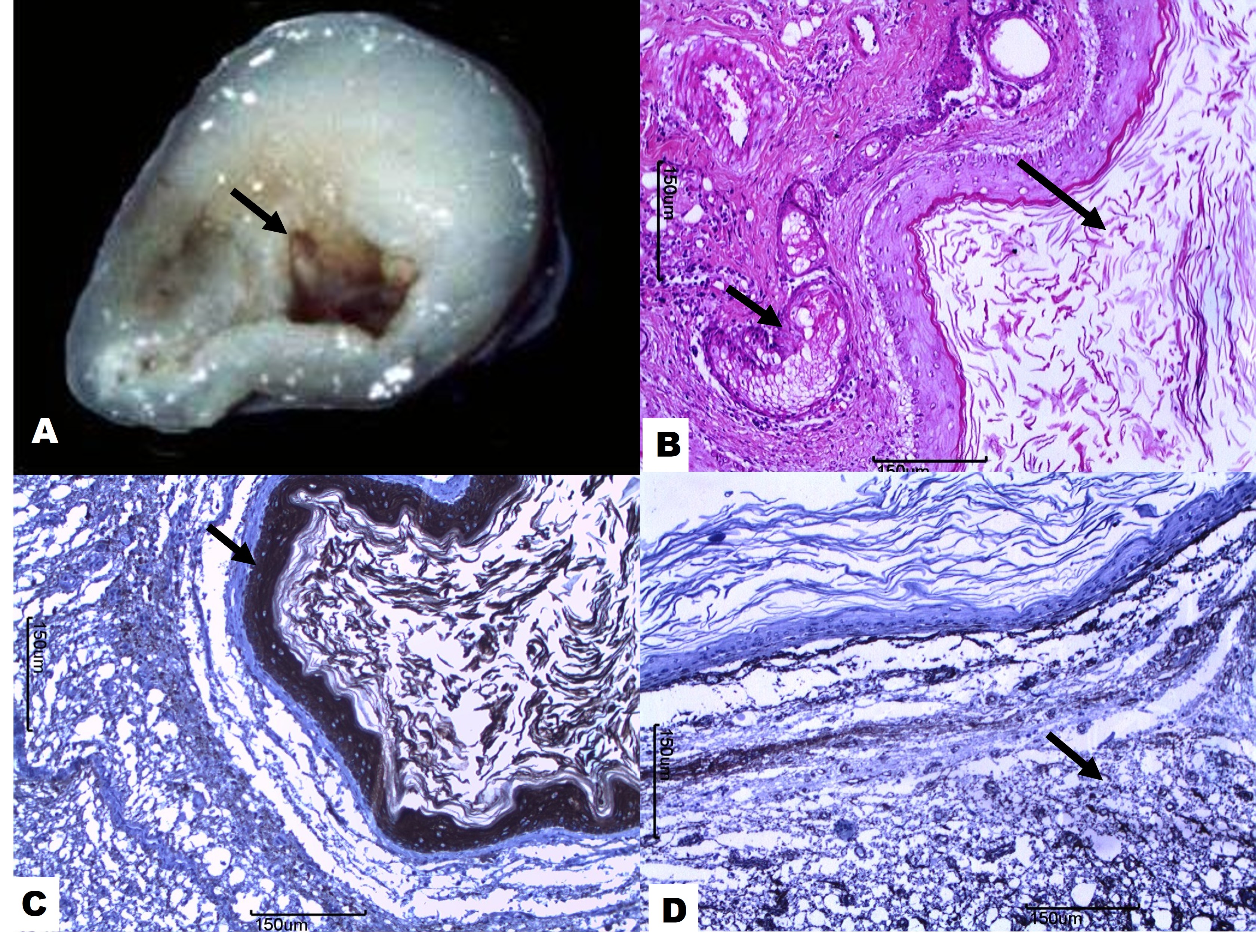
Dermoid cysts are benign congenital lesions of ectodermal origin lined by stratified squamous epithelium and containing cutaneous adnexal structures, keratin debris, and sebaceous material. These lesions are commonly identified in the skin of dogs and cats, particularly along embryonic fusion lines. Occurrence within the central nervous system is uncommon and intramedullary localization is exceptionally rare. This report describes an intramedullary dermoid cyst associated with Wallerian degeneration in an eight-month-old female Boxer dog presenting with a one-month history of paraplegia, self-mutilation of the right pelvic limb paw, and loss of deep pain perception. Due to severe neurological dysfunction, euthanasia was performed and the animal was submitted for pathological examination. Gross examination revealed a 2 cm thin-walled, pearly white cystic lesion located within the L4–S3 spinal cord segments, containing friable brownish material with keratin filaments. Histopathological examination demonstrated a cyst lined by well-differentiated stratified squamous keratinizing epithelium with a prominent stratum granulosum and abundant orthokeratotic laminated keratin within the lumen. Cutaneous adnexal structures, including sebaceous and apocrine glands, were observed within the cyst wall. Adjacent spinal cord tissue exhibited severe compression, focal malacia, and Wallerian degeneration characterized by digestion chambers and gitter cells. Immunohistochemically, the epithelial lining showed strong cytokeratin immunostaining and absence of vimentin expression, whereas stromal and adnexal components were vimentin-positive. The morphological diagnosis was an intramedullary dermoid cyst associated with Wallerian degeneration. This report highlights the rare intramedullary presentation of dermoid cysts in dogs and reinforces the importance of histopathological and immunohistochemical evaluation for definitive diagnosis.
Downloads
Published
Issue
Section
License
Copyright (c) 2026 Brazilian Journal of Veterinary Pathology

This work is licensed under a Creative Commons Attribution 4.0 International License.