
Malignant melanoma in a lamb: clinical and pathological findings
DOI:
https://doi.org/10.24070/bjvp.1983-0246.019020Keywords:
cytology, histopathology, immunohistochemistry, sheep, melanocytes, neoplasiaAbstract
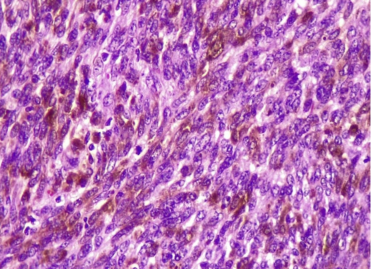
Melanoma is a rare neoplasm in small ruminants, particularly in sheep, and is uncommon in young animals. This study describes the clinical, cytological, histopathological, and immunohistochemical findings of a melanoma in a lamb. A three-month-old female sheep presented with a progressively growing mass in the left periauricular region, first observed at 45 days of age. Cytological evaluation revealed atypical cellular proliferation with marked pleomorphism and mitotic figures, suggesting a malignant neoplastic process. Imaging examinations demonstrated increased soft-tissue volume without evidence of bone involvement. Surgical excision was performed for both diagnostic and therapeutic purposes. Histopathological examination revealed a non-encapsulated, densely cellular, infiltrative neoplasm predominantly composed of epithelioid cells, with marked anisocytosis and anisokaryosis, a high mitotic index, intracytoplasmic melanin pigment, and areas of necrosis. Immunohistochemical analysis showed positive immunostaining for S100 and Melan-A, confirming the melanocytic origin of the neoplasm. Despite surgical removal, rapid local recurrence and clinical deterioration were observed, culminating in death within 30 days, indicating aggressive biological behavior and possible metastatic dissemination. This report demonstrates that, although rare in sheep, melanoma can occur in young animals and may progress rapidly, with a poor prognosis, highlighting the importance of an integrated diagnostic approach and its inclusion in the differential diagnosis of cutaneous masses in lambs.
Downloads
Published
Issue
Section
License
Copyright (c) 2026 Brazilian Journal of Veterinary Pathology

This work is licensed under a Creative Commons Attribution 4.0 International License.